touchOPHTHALMOLOGY touchOPHTHALMOLOGY
Glaucoma
Read Time: 3 mins

Keratoplasty with Glaucoma—Survive or Dive?

Copy Link
Published Online: Mar 9th 2016 US Ophthalmic Review, 2016;9(1):23–4 DOI: http://doi.org/10.17925/USOR.2016.09.01.1b
Authors: Marianne O Price, Francis W Price
Quick Links:
Abstract
Article
Article Information
Abstract:
Overview

Keratoplasty and glaucoma are interconnected: glaucoma can precipitate corneal decompensation and accelerate graft failure, while keratoplasty can complicate intraocular pressure (IOP) control. Advances in endothelial keratoplasty (EK) provide significant benefits for glaucoma patients. Prior filtration surgery remains the greatest risk factor for EK failure - newer micro-invasive techniques may change that.

Keywords

Penetrating keratoplasty, endothelial keratoplasty, glaucoma, aqueous shunt, trabeculectomy, DSEK, DSAEK, DMEK, MIGS

Article:

Prior glaucoma, whether medically or surgically managed, is a significant risk factor for premature failure of penetrating keratoplasty (PK).1 Ten-year PK outcomes show that prior glaucoma significantly increases the risk of all three leading causes of graft failure:

• doubling the risk of failure from rejection;
• doubling the risk of endothelial decompensation in the absence of rejection; and
• tripling the risk of failure from ocular surface complications.1

Fortunately, advances in endothelial keratoplasty (EK) now provide many advantages for glaucoma patients.

Glaucoma is Associated with Graft Failure
The multicenter Cornea Donor Study found that the risk of PK failure was two times higher in eyes with prior medically managed glaucoma, three times higher in eyes with surgically managed glaucoma, and seven times higher with the use of glaucoma medications plus prior glaucoma surgery.2

These results raise the question of why glaucoma is associated with PK failure. The answer is that PK severs corneal nerves, produces an anesthetic cornea, and requires long-standing sutures. The toxic preservatives in glaucoma eye drops further aggravate the disrupted corneal surface after PK, increasing the risk of ocular surface disease, inflammation, and graft rejection. EK, however, is beneficial for patients with medically managed glaucoma, because it is performed with a small incision, maintains corneal sensation and requires little to no suture duration, so it has minimal risk of ocular surface complications and a lower risk of rejection. In fact, the authors have found that EK survival in patients with medically managed glaucoma is nearly as good as in patients without prior glaucoma.3

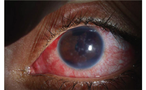
Unfortunately, pseudophakic corneal edema is increasingly associated with previous glaucoma surgery. During the 1980s and 1990s, only 3% of eyes treated by the authors for pseudophakic corneal edema had a prior trabeculectomy or aqueous shunt,4 whereas the proportion was tenfold higher in cases treated between 2003 and 2005 (33% had prior filtration surgery).4

The authors found that prior glaucoma surgery is the single biggest risk factor for EK failure.3 In an analysis of 453 EK recipients, our five-year graft survival rate was 94% in eyes without previous glaucoma, 90% in eyes with previous medically managed glaucoma, 59% in eyes with a prior trabeculectomy, and only 25% in eyes with a prior aqueous shunt.3 Other centers have reported similarly poor graft survival after glaucoma surgery.5,6

Why is prior glaucoma surgery so detrimental to keratoplasty survival? Certainly, a tube that touches the cornea is a risk factor for endothelial decompensation, but the tubes were carefully placed and trimmed to avoid corneal touch, therefore, it is unlikely that this was a significant contributor to graft failure. The authors sampled the aqueous humor from eyes with prior glaucoma surgery and found that the total protein concentration was approximately fivefold higher than normal in eyes with a trabeculectomy and about tenfold higher than normal in eyes with an aqueous shunt (either valved or non-valved).7,8 Additionally, the relative concentrations of hundreds of aqueous humor proteins were significantly altered after filtration surgery,7 suggesting that the aqueous environment was no longer optimal for corneal endothelial survival.

Given these findings, it is important to set realistic expectations for keratoplasty candidates with prior glaucoma surgery. Glaucoma surgery is important because we cannot replace the optic nerve and, while surgery may cause the transplant to fail sooner, it is easy to replace the transplant, especially with EK.

On the flip side of the coin, keratoplasty can cause or exacerbate glaucoma, either because of the changes to the angle sometimes caused by PK, or because of the long-term use of topical corticosteroids to prevent graft rejection. The reported rate of intraocular pressure (IOP) elevation after keratoplasty ranges from 9–35% in eyes without prior glaucoma and from 29–80% in eyes with prior glaucoma.9

Descemet Membrane Endothelial
Keratoplasty Fortunately, rejection rates are lower with EK than PK, and they are remarkably low with the technique known as Descemet membrane endothelial keratoplasty (DMEK), in which only donor endothelium and Descemet membrane are implanted.10 The two-year cumulative risk of a rejection episode is <1% after DMEK,10,11 which is more than tenfold lower than with Descemet stripping endothelial keratoplasty (DSEK) or PK when using the same corticosteroid dosing regimen.10

Prospective, randomized studies have shown that the remarkably low risk of rejection with DMEK makes it possible to use weaker topical corticosteroids and thereby substantially reduce the risk of IOP elevation (by two to three times) without significantly increasing the risk of graft rejection episodes.12,13 At the author’s clinic, steroid strength is reduced at one to two months after DMEK in Caucasians, but a higher steroid strength is maintained for African Americans and those with very darkly pigmented eyes, because they have a higher risk of graft rejection.14

Conclusion
In conclusion, the advent of EK has benefited glaucoma patients, because EK maintains an intact corneal surface, so it is easier to accurately measure vision, IOP and visual fields, and there are few to no ocular surface complications. In addition, the EK technique known as DMEK has a remarkably low risk of rejection, so the steroid strength can be safely reduced to decrease the risk of IOP elevation. Whereas both medically and surgically managed glaucoma adversely impact PK survival, only prior glaucoma filtration surgery significantly reduces EK survival. Keratoplasty candidates with a prior tube or trabeculectomy should be counseled about the potential need to replace the graft sooner than usual. We are hopeful that some of the new micro-invasive glaucoma procedures will prove to be more favorable for keratoplasty survival than traditional tubes and trabs.

Article Information:
Disclosure

Marianne O Price and Francis W Price have received a research grant from Bausch & Lomb and are consultants for Transcend Medical. No funding was received in
the publication of this article. This article is a short opinion piece and has not been submitted to external peer review.

Correspondence

Marianne O Price, PhD, Cornea Research Foundation of America, 9002 North Meridian Street, Suite 212, Indianapolis, IN 46260, US. E: mprice@cornea.org

Access

This article is published under the Creative Commons Attribution Noncommercial License, which permits any noncommercial use, distribution, adaptation, and
reproduction provided the original author(s) and source are given appropriate credit.

Received

2016-02-03T00:00:00

References

1. Price MO, Thompson RW Jr, Price FW Jr, Risk factors for various causes of failure in initial corneal grafts, Arch Ophthalmol, 2003;121:1087–92.
2. Sugar A, Tanner JP, Dontchev M, et al., Recipient risk factors for graft failure in the cornea donor study, Ophthalmology, 2009;116:1023–8.
3. Anshu A, Price MO, Price FW, Descemet’s stripping endothelial keratoplasty: long-term graft survival and risk factors for failure in eyes with preexisting glaucoma, Ophthalmology, 2012;119:1982–7.
4. Price MO, Fairchild KM, Price DA, Price FW Jr, Descemet stripping endothelial keratoplasty five-year graft survival and endothelial cell loss, Ophthalmology, 2011;118:725–9.
5. Aldave AJ, Chen JL, Zaman AS, et al., Outcomes after DSEK in 101 eyes with previous trabeculectomy and tube shunt implantation, Cornea, 2014;33:223–9.
6. Nahum Y, Mimouni M, Busin M, Risk factors predicting the need for graft exchange after Descemet stripping automated endothelial keratoplasty, Cornea, 2015;34:876–9.
7. Rosenfeld M, Price MO, Lai X, et al., Distinctive and pervasive alterations in aqueous humor protein composition following different types of glaucoma surgery, Mol Vision, 2015;21:911–8.
8. Anshu A, Price MO, Richardson MR, et al., Alterations in the aqueous humor proteome in patients with a glaucoma shunt device, Mol Vision, 2011;17:1891–900.
9. Vajaranant TS, Price MO, Price FW, et al., Visual acuity and intraocular pressure after Descemet’s stripping endothelial keratoplasty in eyes with and without preexisting glaucoma, Ophthalmology 2009;116:1644–50.
10. Anshu A, Price MO, Price FW Jr, Risk of corneal transplant rejection significantly reduced with Descemet’s membrane endothelial keratoplasty, Ophthalmology, 2012;119:536–40.
11. Dapena I, Ham L, Netukova M, et al., Incidence of early allograft rejection after Descemet membrane endothelial keratoplasty, Cornea, 2011;30:1341–5.
12. Price MO, Price FW Jr, Kruse FE, et al., Randomized comparison of topical prednisolone acetate 1% versus fluorometholone 0.1% in the first year after Descemet membrane endothelial keratoplasty, Cornea, 2014;33:880–6.
13. Price MO, Feng MT, Scanameo A, et al., Loteprednol etabonate 0.5% gel vs. prednisolone acetate 1% solution after Descemet membrane endothelial keratoplasty: prospective, randomized trial, Cornea, 2015;34:853–8.
14. Price MO, Jordan CS, Moore G, Price FW Jr, Graft rejection episodes after Descemet stripping with endothelial keratoplasty; part two: the statistical analysis of probability and risk factors, Br J Ophthalmol, 2009;93:391–5.

Further Resources

Share this Article
Related Content In Glaucoma
  • Copied to clipboard!
    accredited arrow-down-editablearrow-downarrow_leftarrow-right-bluearrow-right-dark-bluearrow-right-greenarrow-right-greyarrow-right-orangearrow-right-whitearrow-right-bluearrow-up-orangeavatarcalendarchevron-down consultant-pathologist-nurseconsultant-pathologistcrosscrossdownloademailexclaimationfeedbackfiltergraph-arrowinterviewslinkmdt_iconmenumore_dots nurse-consultantpadlock patient-advocate-pathologistpatient-consultantpatientperson pharmacist-nurseplay_buttonplay-colour-tmcplay-colourAsset 1podcastprinter scenerysearch share single-doctor social_facebooksocial_googleplussocial_instagramsocial_linkedin_altsocial_linkedin_altsocial_pinterestlogo-twitter-glyph-32social_youtubeshape-star (1)tick-bluetick-orangetick-red tick-whiteticktimetranscriptup-arrowwebinar Sponsored Department Location NEW TMM Corporate Services Icons-07NEW TMM Corporate Services Icons-08NEW TMM Corporate Services Icons-09NEW TMM Corporate Services Icons-10NEW TMM Corporate Services Icons-11NEW TMM Corporate Services Icons-12Salary £ TMM-Corp-Site-Icons-01TMM-Corp-Site-Icons-02TMM-Corp-Site-Icons-03TMM-Corp-Site-Icons-04TMM-Corp-Site-Icons-05TMM-Corp-Site-Icons-06TMM-Corp-Site-Icons-07TMM-Corp-Site-Icons-08TMM-Corp-Site-Icons-09TMM-Corp-Site-Icons-10TMM-Corp-Site-Icons-11TMM-Corp-Site-Icons-12TMM-Corp-Site-Icons-13TMM-Corp-Site-Icons-14TMM-Corp-Site-Icons-15TMM-Corp-Site-Icons-16TMM-Corp-Site-Icons-17TMM-Corp-Site-Icons-18TMM-Corp-Site-Icons-19TMM-Corp-Site-Icons-20TMM-Corp-Site-Icons-21TMM-Corp-Site-Icons-22TMM-Corp-Site-Icons-23TMM-Corp-Site-Icons-24TMM-Corp-Site-Icons-25TMM-Corp-Site-Icons-26TMM-Corp-Site-Icons-27TMM-Corp-Site-Icons-28TMM-Corp-Site-Icons-29TMM-Corp-Site-Icons-30TMM-Corp-Site-Icons-31TMM-Corp-Site-Icons-32TMM-Corp-Site-Icons-33TMM-Corp-Site-Icons-34TMM-Corp-Site-Icons-35TMM-Corp-Site-Icons-36TMM-Corp-Site-Icons-37TMM-Corp-Site-Icons-38TMM-Corp-Site-Icons-39TMM-Corp-Site-Icons-40TMM-Corp-Site-Icons-41TMM-Corp-Site-Icons-42TMM-Corp-Site-Icons-43TMM-Corp-Site-Icons-44TMM-Corp-Site-Icons-45TMM-Corp-Site-Icons-46TMM-Corp-Site-Icons-47TMM-Corp-Site-Icons-48TMM-Corp-Site-Icons-49TMM-Corp-Site-Icons-50TMM-Corp-Site-Icons-51TMM-Corp-Site-Icons-52TMM-Corp-Site-Icons-53TMM-Corp-Site-Icons-54TMM-Corp-Site-Icons-55TMM-Corp-Site-Icons-56TMM-Corp-Site-Icons-57TMM-Corp-Site-Icons-58TMM-Corp-Site-Icons-59TMM-Corp-Site-Icons-60TMM-Corp-Site-Icons-61TMM-Corp-Site-Icons-62TMM-Corp-Site-Icons-63TMM-Corp-Site-Icons-64TMM-Corp-Site-Icons-65TMM-Corp-Site-Icons-66TMM-Corp-Site-Icons-67TMM-Corp-Site-Icons-68TMM-Corp-Site-Icons-69TMM-Corp-Site-Icons-70TMM-Corp-Site-Icons-71TMM-Corp-Site-Icons-72