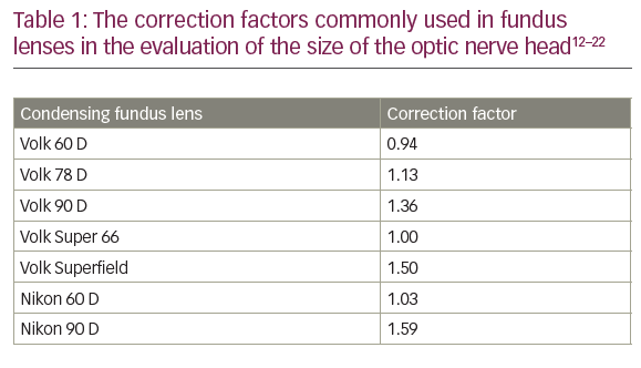
Canon is a world leader and innovator of imaging and information technology solutions. Its dedication to breaking new ground has been continuously demonstrated during the company’s history, in particular with the birth of Canon’s electro-optical system (EOS) single lens reflex (SLR) technology more than two decades ago, which has since given rise to a range of revolutionary imaging and photographic solutions for multiple applications.
For example, 2005 marked the launch of the EOS 20Da, a camera that was designed exclusively and uniquely for astrophotography. The design of the EOS 20Da was based on the Canon EOS 20D camera, but was modified to cater for the exacting needs of photographers specialising in astrophotography. For example, the EOS 20Da infrared-blocking filter allowed the passage of 2.5 times more light than the EOS 20D at a wavelength of around 656nm for enhanced detail and transmission of red light – just one factor that made the camera popular among the astrophotography community and a true innovation of its time.
In line with this trend, Canon has been pioneering improvements in healthcare and medical diagnosis for nearly 70 years, and today it is a leading provider of eye-care imaging solutions.
Incorporating Canon’s latest EOS digital SLR technology for excellent imaging quality, fully optimised for multiple-mode retinal photography, the Canon CX-1 digital retinal camera is the latest addition to its eyecare portfolio and a true innovation in digital retinal imaging.
A fully hybrid digital retinal camera synergised for digital mydriatic and non-mydriatic imaging, the Canon CX-1 is an easy-to-use, compact and fully portable system that is capable of capturing highresolution images in multiple modes, without the need for additional cameras or a monitor. As a result, users of the CX-1 can deliver convenient, comfortable procedures to their patients while expanding their range of diagnostic capabilities.
The CX-1 captures high-quality images in five photography modes: colour, red-free, cobalt, fluorescein angiography and even technically challenging fundus autofluorescence (FAF). Moreover, users can capture these images in mydriatic or non-mydriatic mode, simply by pushing a single button to switch between modes.
Canon’s EOS digital SLR technology, with its renowned imageprocessing capabilities, has been adapted exclusively for use within CX-1; its proprietary 15.1-megapixel complementary metal oxide semiconductor (CMOS) sensor assures high-quality images in all modes.
Meanwhile, the liquid crystal display (LCD) monitor handles magnification, focus and alignment, and even provides stereo guides for successful image capture and management.
Another important feature is the EOS camera’s internal digital imaging integrated circuit (DIGIC) processor, which allows high-speed processing as an extension of the CX-1 retinal imaging control software for increased efficiency of image delivery for review in spite of the highresolution of imagery. It even improves noise reduction in high ISO images as well as effectively compensating for interference from cataracts, allowing optimal image quality during all FAF photography.
The system itself has been ergonomically designed and so is compact and portable for superior convenience, ease of use and patient comfort.
The CX-1 combines Canon’s long-standing history of innovation in retinal imaging with the Canon EOS digital SLR camera technology to create a truly innovative product.