touchOPHTHALMOLOGY touchOPHTHALMOLOGY
Anterior Segment, Corneal and External Disorders
Read Time: 3 mins

Preserved Amniotic Membrane Transplantation for Ocular Surface Reconstruction

Copy Link
Published Online: Feb 18th 2011 US Ophthalmic Review, 2007,3:35-7 DOI: http://doi.org/10.17925/USOR.2007.03.00.35
Authors: Alisa Kim, Roy S Chuck
Quick Links:
Article
Article Information
Article:

Maintenance of the ocular surface is dependent on the complex interplay of the lids, lacrimal gland, tear film, conjunctiva, cornea, and neural network. Although multifactorial in etiology, severe ocular surface disorders (OSDs) commonly result in progressive inflammation, vascularization, scarring, and/or loss of visual function, often refractory to conservative medical therapy. Unique properties of human amniotic membrane (AM) confer several advantages in the management and treatment of various OSDs. AM is the innermost layer of the placenta and is composed of an epithelial monolayer, an underlying thick basement membrane, and an avascular stroma.

The first ophthalmic utilization of fresh placental tissue by de Rötth was reported in 1940 with limited success.1 Since then, AM transplantation (AMT) has been used successfully as a temporary patch and/or permanent graft for corneal and/or conjunctival cells in the management of numerous conditions including persistent epithelial defects, corneal ulcers, descemetocoele formation, corneal perforation, limbal stem cell deficiency, symptomatic bullous keratopathy, band keratopathy, chemical injury, thermal injury, repair of conjunctival defects, pterygium surgery, bleb repair, scleral perforation, and high-risk corneal transplantation.

Although the exact mechanism of action is unclear, AM demonstrates physiological properties that promote epithelial and stromal wound healing while suppressing inflammation, fibrosis, and vascularization. The AM basement membrane contains laminin, fibronectin, and collagen type IV and VII, similar to the basement membrane of conjunctival tissue, but more than corneal epithelium.2–5 The basement membrane provides a substrate that promotes epithelial cell adhesion, migration, and differentiation5–7 and prevents epithelial cell apoptosis.8,9 The underlying stromal matrix contains proteins that suppress transforming growth factor (TGF)-β signaling in fibroblasts, and consequently inhibit the proliferation and differentiation into myofibroblasts, extracellular matrix production, and scar formation. The resultant downregulation is responsible for the antifibrosis effect.10–12

The anti-inflammatory activity of AM is attributed to numerous mediators, including interleukin (IL)-1 receptor antagonist, IL-10, activin, inhibin, and inter-α-trypsin inhibitor.12–14 AM has also been shown to induce apoptosis of inflammatory cells.14–17 Vascularization is inhibited by the antiinflammatory effect of AM and by the antiangiogenic proteins present within AM, including endostatin, thrombospondin-1, and inhibitors of metallo-proteases.13,18 Finally, a unique advantage of AM is the low immunogenicity of the tissue and, therefore, the consequent lack of rejection.4,19–21 The immunosuppressive ability of AM to inhibit a mixed lymphocyte reaction appears to be partly mediated by a soluble factor, resulting in decreased alloreactive T-cell proliferation and Th1 and Th2 cytokine production.

Advances in technology have allowed surgeons to move away from the disadvantages and inconveniences of fresh AM to cryopreserved, dehydrated, or freeze-dried AM tissue. In the US, the two different AMs are commercially available as epithelialized, cryopreserved AM (Amniograft®, Biotissue Inc., Miami, Florida) or de-epithelialized, dehydrated AM (AmbioDry™, IOP, Inc., Costa Mesa, California). AM is obtained under sterile conditions after elective Cesarean section. The cryopreserved AM contains epithelial cells and is disinfected with antimicrobial agents, attached to nitrocellulose paper with the basement membrane facing upward, stored in a deep freeze at -80°C, and thawed for five to 10 minutes before use. Although the AM is not sterilized during its processing, the tissue undergoes microbiological testing prior to its release. The orientation is distinguishable by the sticky stromal side attached to the nitrocellulose paper. When stored properly at -80°C, the membrane can be used for up to two years from the time of procurement. The dehydrated AM undergoes de-epithelialization, dehydration, sterilization with electron beam irradiation, and storage free-standing at room temperature (50–85°F). As free-standing tissue, the AM does not require careful separation from nitrocellulose paper, and the orientation is clear by the imprinted text ‘IOP.’ In the dry state, the AM can be cut, manipulated, and placed onto the surgical site before being activated with a saline solution or an adhesive agent. The AM can be used for up to two years from the time of processing. Compared with AM with epithelial cells, AM denuded of epithelial cells may provide a better substrate for the culturing of corneal epithelial cells,22 although this study was not performed with conjunctival epithelial cells. A study by Chuck et al. that evaluated the biomechanical properties of AM showed that the cryopreserved AM tolerated a higher maximum stress prior to rupture and stretched more than twice the length of the dehydrated AM, suggestive of greater elasticity.23 There have been no comparative clinical studies evaluating the efficacy of cryopreserved and dehydrated AMT in the management of OSDs.

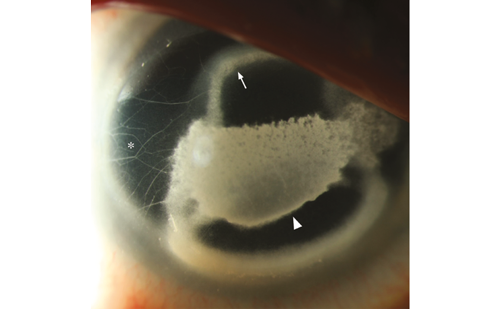
In the field of ocular surface reconstruction, the use of AMT in the surgical management of pterygium has gained popularity since the first utilization by Prabhasawat in 1997.24 In addition to the surgical goals of successful removal of the pterygium and achieving an optimal postoperative cosmetic result, the primary concern for the surgeon is recurrence. The clinical results with AMT have been variable, with recurrence rates ranging from 3 to 41%24–31 for primary pterygium and from 9.5 to 52.6% for recurrent pterygium.24,27,30–33 Although the rate of recurrence with AMT is much lower than with primary closure, the results compared with conjunctival autografting alone have not been consistent.24–26,28,30,3127 and adjunctive injection of corticosteroid,27,34 have been associated with lower recurrence rates. No significant difference in rate of recurrence was found with AMT combined with intraoperative mitomycin C (MMC).32 The combination of a conjunctival and/or limbal autograft33,35–37 with AMT facilitates a more rapid re-population with normal epithelial cells, which may decrease inflammation and possibly contribute a barrier effect against fibrovascular invasion. Difficult cases may benefit from simultaneous AMT, conjunctival limbal autograft, and MMC application,38–40 particularly multirecurrent cases.

The key to successful treatment centers on suppression of inflammation. Pro-inflammatory cytokines (IL-1β, tumor necrosis factor [TNF]-α) can promote cultured fibroblast proliferation41 and overexpression of select matrix metalloproteases (type I, III), which allows for a more invasive cellular phenotype42,43 that could increase the likelihood of recurrence. Therefore, the potential anti-inflammatory, antifibrotic, and antiangiogenic benefits of AM may provide several advantages in ocular surface reconstruction after pterygium excision. Using indocyanine green angiography (ICGA), delayed vascularization in the AMT graft has been compared with conjunctival autograft transplantation (CAT), which may be attributable to the antiangiogenic effects of AM.44 These findings may contribute to the delay in recurrence with AMT compared with limbal CAT (LCAT), which has been postulated as a barrier phenomenon of AMT.45 In addition to the inherent physiological properties of AMT, other benefits include convenience, less technical skill needed, shorter operative time, and fewer complications compared with harvesting a conjunctival (and/or limbal) graft. Another distinct advantage of AMT is the ability to preserve conjunctival tissue in precluding the use of a CAT, particularly for future glaucoma-filtering procedures. AM grafting is considered the preferred adjunctive treatment in large pterygia where insufficient conjunctiva for a graft is available or in cases that are at high risk for recurrence, as AM transplantation can be repeated.

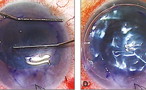
Another technological innovation that has furthered the use of AMT in pterygium surgery is the development of biological tissue adhesives, such as fibrin glue (FG) (Tisseel VH fibrin sealant, Baxter Corporation, Irvine, California). A tissue bioadhesive can be used to supplement sutures to reduce the number needed or can be used in place of sutures. The initial success of FG in conjunction with CAT46–49,50 led to an increased interest in AM grafts. Implementation of FG to secure the AM graft compared with sutures has been associated with shorter surgical times, technical ease of use, and less post-operative discomfort, particularly foreign body sensation.46–49,51 The decreased operative time facilitates the use of topical anesthesia. Additionally, FG avoids the suture-related complications of globe perforation, hemorrhage, graft button-hole, suture abscess, tissue necrosis, and giant papillary conjunctivitis and bypasses the need to remove sutures post-operatively, as the fibrin naturally biodegrades. Lower rates of recurrence have been found for FG (5.3–8%) compared with sutures (13.5–20%) to secure conjunctival autografts in pterygium surgery.48,50 Kheirkhah et al. found significantly greater conjunctival inflammation after AMT and MMC with sutures compared with FG.52 More severe inflammation has been found to be related to higher rates of recurrence.52,53

AM has been modified to broaden its utility. Minimal amounts of FG are recommended due to its potential pro-inflammatory effect. To address this concern a bio-adhesive-coated AMT (fibrinogen and thrombin) was created, maintaining the biological characteristics of AM and bypassing the need for sutures.54 In addition to use as a graft, AM has been used as a temporary patch for the surgical treatment of primary treatment without recurrence (n=20).55 In conjunction with a fixed AMT graft, a temporary AMT patch may be beneficial after pterygium surgery, similar to sutureless AMT for partial limbal stem cell deficiency.56 AM is commercially available to be used as a temporary patch with cryopreserved AM fastened to a symblepharon ring (ProKera®, Bio-Tissue Inc, Miami). Other modifications to AMT include ex vivo epithelial cell expansion, which is beyond the scope of this article.

The literature to support the utility of AMT in ocular surface reconstruction continues to expand. Prospective clinical studies are needed to evaluate the efficacy of AMT in comparison with currently available alternatives. Additionally, research to better elucidate the mechanism of action behind the physiological properties of AMT continues to be of great interest. Although limitations of AMT exist, particularly in cases of total stem cell deficiency and severe keratoconjunctivitis sicca, we can expect that future modifications in surgical technique, concomitant medical therapy, and technological advances will develop to optimize the results of procedures using AMT.

Article Information:
Received

2011-02-18T00:00:00

References

  1. de Rötth A, Plastic repair of conjunctival defects with fetal membrane, Arch Ophthalmol, 1940;23:522–5.
  2. Fukuda K, Chikama T, Nakamura M, Nishida T, Differential distribution of subchains of the basement membrane components type IV collagen and laminin among the amniotic membrane, cornea, and conjunctiva, Cornea, 1999;18(1):73–9.
  3. Endo K, Nakamura T, Kawasaki S, Kinoshita S, Human amniotic membrane, like corneal epithelial basement membrane, manifests the alpha5 chain of type IV collagen, Invest Ophthalmol Vis Sci, 2004;45(6):1771–4.
  4. Nakamura T, Yoshitani M, Rigby H, et al., Sterilized, freeze-dried amniotic membrane: A useful substrate for ocular surface reconstruction, Invest Ophthalmol Vis Sci, 2004;45(1):93–9.
  5. Nakamura T, Sekiyama E, Takaoka M, et al., The use of trehalosetreated freeze-dried amniotic membrane for ocular surface reconstruction, Biomaterials, 2008;29(27):3729–37.
  6. Grueterich M, Tseng SC, Human limbal progenitor cells expanded on intact amniotic membrane ex vivo, Arch Ophthalmol, 2002; 120(6):783–90.
  7. Meller D, Tseng SC, Conjunctival epithelial cell differentiation on amniotic membrane, Invest Ophthalmol Vis Sci, 1999;40(5): 878–86.
  8. Boudreau N, Sympson CJ,Werb Z, Bissell MJ, Suppression of ICE and apoptosis in mammary epithelial cells by extracellular matrix, Science, 1995;267(5199):891–3.
  9. Boudreau N,Werb Z, Bissell MJ, Suppression of apoptosis by basement membrane requires three-dimensional tissue organization and withdrawal from the cell cycle, Proc Natl Acad Sci U S A, 1996;93(8):3509–13.
  10. Tseng SC, Li DQ, Ma X, Suppression of transforming growth factor-beta isoforms, TGF-beta receptor type II, and myofibroblast differentiation in cultured human corneal and limbal fibroblasts by amniotic membrane matrix, J Cell Physiol, 1999;179(3): 325–35.
  11. Lee SB, Li DQ, Tan DT, et al., Suppression of TGF-beta signaling in both normal conjunctival fibroblasts and pterygial body fibroblasts by amniotic membrane, Curr Eye Res, 2000;20(4): 325–34.
  12. Tseng SC, Espana EM, Kawakita T, et al., How does amniotic membrane work?, Ocul Surf, 2004;2(3):177–87.
  13. Hao Y, Ma DH, Hwang DG,et al., Identification of antiangiogenic and antiinflammatory proteins in human amniotic membrane, Cornea, 2000;19(3):348–52.
  14. Solomon A, Rosenblatt M, Monroy D, et al., Suppression of interleukin 1alpha and interleukin 1beta in human limbal epithelial cells cultured on the amniotic membrane stromal matrix, Br J Ophthalmol, 2001;85(4):444–9.
  15. Shimmura S, Shimazaki J, Ohashi Y, Tsubota K, Antiinflammatory effects of amniotic membrane transplantation in ocular surface disorders, Cornea, 2001;20(4):408–13.
  16. Wang MX, Gray TB, Park WC, et al., Reduction in corneal haze and apoptosis by amniotic membrane matrix in excimer laser photoablation in rabbits, J Cataract Refract Surg, 2001;27(2): 310–19.
  17. Li W, He H, Kawakita T, et al., Amniotic membrane induces apoptosis of interferon-gamma activated macrophages in vitro, Exp Eye Res, 2006;82(2):282–92.
  18. Kim JC, Tseng SC, The effects on inhibition of corneal neovascularization after human amniotic membrane transplantation in severely damaged rabbit corneas, Korean J Ophthalmol, 1995;9(1):32–46.
  19. Adinolfi M, Akle CA, McColl I, et al., Expression of HLA antigens, beta 2-microglobulin and enzymes by human amniotic epithelial cells, Nature, 1982;295(5847):325–7.
  20. Akle CA, Adinolfi M,Welsh KI, et al., Immunogenicity of human amniotic epithelial cells after transplantation into volunteers, Lancet, 1981;2(8254):1003–5.
  21. Kubo M, Sonoda Y, Muramatsu R, Usui M, Immunogenicity of human amniotic membrane in experimental xenotransplantation, Invest Ophthalmol Vis Sci, 2001;42(7):1539–46.
  22. Koizumi N, Fullwood NJ, Bairaktaris G, et al., Cultivation of corneal epithelial cells on intact and denuded human amniotic membrane, Invest Ophthalmol Vis Sci, 2000;41(9):2506–13.
  23. Chuck RS, Graff JM, Bryant MR, Sweet PM, Biomechanical characterization of human amniotic membrane preparations for ocular surface reconstruction, Ophthalmic Res, 2004;36(6):341–8.
  24. Prabhasawat P, Barton K, Burkett G, Tseng SC, Comparison of conjunctival autografts, amniotic membrane grafts, and primary closure for pterygium excision, Ophthalmology, 1997;104(6): 974–85.
  25. Tananuvat N, Martin T, The results of amniotic membrane transplantation for primary pterygium compared with conjunctival autograft, Cornea, 2004;23(5):458–63.
  26. Ma DH, See LC, Liau SB, Tsai RJ, Amniotic membrane graft for primary pterygium: Comparison with conjunctival autograft and topical mitomycin C treatment, Br J Ophthalmol, 2000;84(9): 973–8.
  27. Solomon A, Pires RT, Tseng SC, Amniotic membrane transplantation after extensive removal of primary and recurrent pterygia, Ophthalmology, 2001;108(3):449–60.
  28. Memarzadeh F, Fahd AK, Shamie N, Chuck RS, Comparison of deepithelialized amniotic membrane transplantation and conjunctival autograft after primary pterygium excision, Eye, 2008;22(1):107–12.
  29. Fernandes M, Sangwan VS, Bansal AK, et al., Outcome of pterygium surgery: Analysis over 14 years, Eye, 2005;19(11): 1182–90.
  30. Luanratanakorn P, Ratanapakorn T, Suwan-Apichon O, Chuck RS, Randomised controlled study of conjunctival autograft versus amniotic membrane graft in pterygium excision, Br J Ophthalmol, 2006;90(12):1476–80.
  31. Kucukerdonmez C, Akova YA, Altinors DD, Comparison of conjunctival autograft with amniotic membrane transplantation for pterygium surgery: Surgical and cosmetic outcome, Cornea, 2007;26(4):407–13.
  32. Ma DH, See LC, Hwang YS,Wang SF, Comparison of amniotic membrane graft alone or combined with intraoperative mitomycin C to prevent recurrence after excision of recurrent pterygia, Cornea, 2005;24(2):141–50.
  33. Shimazaki J, Shinozaki N, Tsubota K, Transplantation of amniotic membrane and limbal autograft for patients with recurrent pterygium associated with symblepharon, Br J Ophthalmol, 1998; 82(3):235–40.
  34. Paris Fdos S, de Farias CC, Melo GB, et al., Postoperative subconjunctival corticosteroid injection to prevent pterygium recurrence, Cornea, 2008;27(4):406–10.
  35. Shimazaki J, Kosaka K, Shimmura S, Tsubota K, Amniotic membrane transplantation with conjunctival autograft for recurrent pterygium, Ophthalmology, 2003;110(1):119–24.
  36. Fallah MR, Golabdar MR, Amozadeh J, et al., Transplantation of conjunctival limbal autograft and amniotic membrane vs mitomycin C and amniotic membrane in treatment of recurrent pterygium, Eye, 2008;22(3):420–24.
  37. Xi XH, Jiang DY, Tang LS, Transplantation of amniotic membrane and amniotic membrane combined with limbal autograft for patients with complicated pterygium, Hunan Yi Ke Da Xue Xue Bao, 2003;28(2):149–51.
  38. Yao YF, Qiu WY, Zhang YM, Tseng SC, Mitomycin C, amniotic membrane transplantation and limbal conjunctival autograft for treating multirecurrent pterygia with symblepharon and motility restriction, Graefes Arch Clin Exp Ophthalmol, 2006;244(2): 232–6.
  39. Sangwan VS, Murthy SI, Bansal AK, Rao GN, Surgical treatment of chronically recurring pterygium, Cornea, 2003;22(1):63–5.
  40. Miyai T, Hara R, Nejima R, et al., Limbal allograft, amniotic membrane transplantation, and intraoperative mitomycin C for recurrent pterygium, Ophthalmology, 2005;112(7):1263–7.
  41. Cunliffe IA, Richardson PS, Rees RC, Rennie IG, Effect of TNF, IL-1, and IL-6 on the proliferation of human tenon's capsule fibroblasts in tissue culture, Br J Ophthalmol, 1995;79(6):590–95.
  42. Solomon A, Li DQ, Lee SB, Tseng SC, Regulation of collagenase, stromelysin, and urokinase-type plasminogen activator in primary pterygium body fibroblasts by inflammatory cytokines, Invest Ophthalmol Vis Sci, 2000;41(8):2154–63.
  43. Li DQ, Lee SB, Gunja-Smith Z, et al., Overexpression of collagenase (MMP-1) and stromelysin (MMP-3) by pterygium head fibroblasts, Arch Ophthalmol, 2001;119(1):71–80.
  44. Kucukerdonmez C, Akova YA, Altinors DD, Vascularization is more delayed in amniotic membrane graft than conjunctival autograft after pterygium excision, Am J Ophthalmol, 2007;143(2):245–9.
  45. Tananuvat N, Martin T, The results of amniotic membrane transplantation for primary pterygium compared with conjunctival autograft, Cornea, 2004;23(5):458–63.
  46. Bahar I,Weinberger D, Dan G, Avisar R, Pterygium surgery: Fibrin glue versus vicryl sutures for conjunctival closure, Cornea, 2006; 25(10):1168–72.
  47. Cohen RA, McDonald MB, Fixation of conjunctival autografts with an organic tissue adhesive, Arch Ophthalmol, 1993;111(9): 1167–8.
  48. Koranyi G, Seregard S, Kopp ED, Cut and paste: A no suture, small incision approach to pterygium surgery, Br J Ophthalmol, 2004; 88(7):911–14.
  49. Marticorena J, Rodriguez-Ares MT, Tourino R, et al., Pterygium surgery: Conjunctival autograft using a fibrin adhesive, Cornea, 2006;25(1):34–6.
  50. Koranyi G, Seregard S, Kopp ED, The cut-and-paste method for primary pterygium surgery: Long-term follow-up, Acta Ophthalmol Scand, 2005;83(3):298–301.
  51. Jain AK, Bansal R, Sukhija J, Human amniotic membrane transplantation with fibrin glue in management of primary pterygia: A new tuck-in technique, Cornea, 2008;27(1):94–9.
  52. Kheirkhah A, Casas V, Sheha H, et al., Role of conjunctival inflammation in surgical outcome after amniotic membrane transplantation with or without fibrin glue for pterygium, Cornea, 2008;27(1):56–63.
  53. Ti SE, Tseng SC, Management of primary and recurrent pterygium using amniotic membrane transplantation, Curr Opin Ophthalmol, 2002;13(4):204–12.
  54. Sekiyama E, Nakamura T, Kurihara E, et al., Novel sutureless transplantation of bioadhesive-coated, freeze-dried amniotic membrane for ocular surface reconstruction, Invest Ophthalmol Vis Sci, 2007;48(4):1528–34.
  55. Ye J, Kook KH, Yao K, Temporary amniotic membrane patch for the treatment of primary pterygium: Mechanisms of reducing the recurrence rate, Graefes Arch Clin Exp Ophthalmol, 2006;244(5): 583–8.
  56. Kheirkhah A, Casas V, Raju VK, Tseng SC, Sutureless amniotic membrane transplantation for partial limbal stem cell deficiency, Am J Ophthalmol, 2008;145(5):787–94.

Further Resources

Share this Article
Related Content In Corneal and External Disorders
  • Copied to clipboard!
    accredited arrow-down-editablearrow-downarrow_leftarrow-right-bluearrow-right-dark-bluearrow-right-greenarrow-right-greyarrow-right-orangearrow-right-whitearrow-right-bluearrow-up-orangeavatarcalendarchevron-down consultant-pathologist-nurseconsultant-pathologistcrosscrossdownloademailexclaimationfeedbackfiltergraph-arrowinterviewslinkmdt_iconmenumore_dots nurse-consultantpadlock patient-advocate-pathologistpatient-consultantpatientperson pharmacist-nurseplay_buttonplay-colour-tmcplay-colourAsset 1podcastprinter scenerysearch share single-doctor social_facebooksocial_googleplussocial_instagramsocial_linkedin_altsocial_linkedin_altsocial_pinterestlogo-twitter-glyph-32social_youtubeshape-star (1)tick-bluetick-orangetick-red tick-whiteticktimetranscriptup-arrowwebinar Sponsored Department Location NEW TMM Corporate Services Icons-07NEW TMM Corporate Services Icons-08NEW TMM Corporate Services Icons-09NEW TMM Corporate Services Icons-10NEW TMM Corporate Services Icons-11NEW TMM Corporate Services Icons-12Salary £ TMM-Corp-Site-Icons-01TMM-Corp-Site-Icons-02TMM-Corp-Site-Icons-03TMM-Corp-Site-Icons-04TMM-Corp-Site-Icons-05TMM-Corp-Site-Icons-06TMM-Corp-Site-Icons-07TMM-Corp-Site-Icons-08TMM-Corp-Site-Icons-09TMM-Corp-Site-Icons-10TMM-Corp-Site-Icons-11TMM-Corp-Site-Icons-12TMM-Corp-Site-Icons-13TMM-Corp-Site-Icons-14TMM-Corp-Site-Icons-15TMM-Corp-Site-Icons-16TMM-Corp-Site-Icons-17TMM-Corp-Site-Icons-18TMM-Corp-Site-Icons-19TMM-Corp-Site-Icons-20TMM-Corp-Site-Icons-21TMM-Corp-Site-Icons-22TMM-Corp-Site-Icons-23TMM-Corp-Site-Icons-24TMM-Corp-Site-Icons-25TMM-Corp-Site-Icons-26TMM-Corp-Site-Icons-27TMM-Corp-Site-Icons-28TMM-Corp-Site-Icons-29TMM-Corp-Site-Icons-30TMM-Corp-Site-Icons-31TMM-Corp-Site-Icons-32TMM-Corp-Site-Icons-33TMM-Corp-Site-Icons-34TMM-Corp-Site-Icons-35TMM-Corp-Site-Icons-36TMM-Corp-Site-Icons-37TMM-Corp-Site-Icons-38TMM-Corp-Site-Icons-39TMM-Corp-Site-Icons-40TMM-Corp-Site-Icons-41TMM-Corp-Site-Icons-42TMM-Corp-Site-Icons-43TMM-Corp-Site-Icons-44TMM-Corp-Site-Icons-45TMM-Corp-Site-Icons-46TMM-Corp-Site-Icons-47TMM-Corp-Site-Icons-48TMM-Corp-Site-Icons-49TMM-Corp-Site-Icons-50TMM-Corp-Site-Icons-51TMM-Corp-Site-Icons-52TMM-Corp-Site-Icons-53TMM-Corp-Site-Icons-54TMM-Corp-Site-Icons-55TMM-Corp-Site-Icons-56TMM-Corp-Site-Icons-57TMM-Corp-Site-Icons-58TMM-Corp-Site-Icons-59TMM-Corp-Site-Icons-60TMM-Corp-Site-Icons-61TMM-Corp-Site-Icons-62TMM-Corp-Site-Icons-63TMM-Corp-Site-Icons-64TMM-Corp-Site-Icons-65TMM-Corp-Site-Icons-66TMM-Corp-Site-Icons-67TMM-Corp-Site-Icons-68TMM-Corp-Site-Icons-69TMM-Corp-Site-Icons-70TMM-Corp-Site-Icons-71TMM-Corp-Site-Icons-72