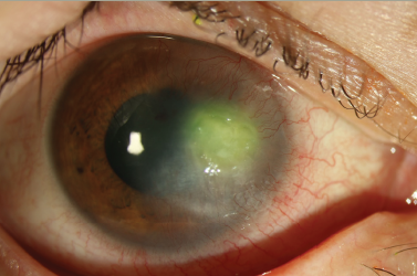
In recent years, reduced corneal biomechanics was identified as an important element in the pathogenesis of various corneal diseases. The biomechanical characteristics of a connective tissue such as strength and resistance against mechanical stress are indispensable prerequisites to maintaining regular shape and function. Intra- and intermolecular cross-links between collagen molecules are essential elements of these biomechanical properties.1–3 Accordingly, collagen cross-links occur physiologically in all organs and tissues with certain biomechanical characteristics.
Cross-linking has been used for decades in various surgical fields to increase the biomechanical properties of connective tissue structures. For example, in cardiac surgery porcine aortic valve bioprostheses are treated with glutaraldehyde prior to implantation to ensure increased cross-linking, which increases biomechanical resistance against biodegradation. Additionally, in ear, nose and throat (ENT) surgery, polymers inducing cross-linking are used in the treatment of destabilised vocal cords and for nasal reconstruction.4–9
In the cornea, a variety of conditions such as primary acquired (keratoconus and pellucid marginal corneal degeneration) and secondary induced (iatrogenic keratectasia after refractive laser surgery) ectatic disorders lead to reduced biomechanical resistance. Corneal collagen cross-linking (CXL) with riboflavin/ ultraviolet A (UVA) represents a new approach to these diseases. To assist researchers and clinicians interested in the field, this article attempts to provide a structured overview of the current state of the method, its basic principles, the technique and its application in primary and iatrogenic keratectasia. Furthermore, it addresses safety issues and potential complications of the method.
The Principle of Corneal Collagen Cross-linking
Inter- and intrafibrillar collagen cross-links are a major factor ensuring the mechanical stiffness of connective tissues. A number of techniques can be used to induce additional cross-links: exposure to aldehydes (glutaraldehyde or aldehyde sugars), enzymatic treatment (lysyloxydase) and photo-polymerisation using UV light.10 For the transparent cornea, some approaches were either too toxic (exposure to glutaraldehyde or UVA alone) or too time-consuming (ribose 0.5 molar solution for 14 days).11,12
The most promising technique was photo-polymerisation by generation of free radicals using a non-toxic and soluble photomediator. This photomediator should absorb UVA light trongly enough to generate free radicals that induce cross-links and protect deeper ocular structures from the potential hazards of free radical formation. These parameters were achieved by using a 0.1% aqueous solution of riboflavin–phosphate (vitamin B2). It provides an adequate shielding effect at a wavelength of 370nm, and generates sufficient free radicals to induce formation of additional collagen cross-links. These cross-links are intramolecular rather than intermolecular, as suggested by an increase in collagen fibre diameter by approximately 10% following CXL.13 This increase was more pronounced in the anterior stroma.13 Whether interfibrillar cross-links are also induced is still unclear.
To reduce uncontrolled stromal swelling, riboflavin solution was diluted in a carrier that was iso-osmolar to the corneal stroma (dextrane 20%). Since riboflavin is a macromolecule (molecular weight 376.37g/mol), the corneal epithelium represents a barrier that decreases the absorption rate.14 The corneal epithelium should therefore be removed prior to instillation of riboflavin solution. The intensity of UVA irradiation was set to 3mW/cm2, corresponding to a surface dose of 5.4J/cm2. These parameters induce corneal crosslinks to a depth of 310μm. A stromal thickness of at least 400μm should be respected in CXL; at this depth, the irradiation intensity is two times lower than the damage threshold level.15–17
CXL leads to a marked increase of corneal biomechanical stability. To determine the stiffening effect, the key experiment is the stress– strain measurement performed in a micro-material tester using corneal strips.18–21 In human cadaver corneas, Young’s modulus increased by a factor of 4.5 using the standard parameters.19
In addition to the biomechanical effect, a biochemical effect contributes to the increased resistance of cross-linked corneas. Spoerl and colleagues have experimentally investigated the effect of enzymatic digestion on porcine corneas in a solution containing pepsin, trypsin and collagenase with and without precedent CXL. The cross-linked corneas were markedly more resistant to the proteolytic process. Changes in the tertiary structure of the collagen molecule may explain the stabilising biochemical effect of cross-linking. These changes prevent proteolytic enzymes from accessing their specific cleavage sites by steric hindrance.22
Wollensak and co-workers studied the cytotoxicity of the riboflavin/ UVA standard treatment (for parameters see below) on keratocytes and endothelial cells.15,16,23 In rabbit corneas, keratocyte apoptosis was detected at a depth of up to 300μm at 24 hours following standard CXL treatment. Smaller irradiances led to shallower cell depth following Lambert-Beer’s law.23 In cell cultures established from porcine keratocytes, the damage threshold of the irradiance of UVA in combination with 0.025% riboflavin solution was determined at 0.45mW/cm2, which is 10 times lower than for UVA irradiation alone.16 A similar experimental set-up was used to measure the damage threshold for porcine endothelial cells.15 At an irradiance of 0.3mW/cm2 no signs of cell damage were detected, whereas at 0.35mW/cm2 98% of the cells stained positively for both trypan blue and yopro in their nuclei. The authors concluded that when using the standard riboflavin/UVA technique, a minimal pre-operative corneal thickness of 400μm after removal of the epithelium is mandatory to avoid damage to the corneal endothelium.
Treating Corneas with a Pre-operative Thickness >400μm (Standard Technique)
The following treatment parameters are currently widely used to treat corneas thicker than 400μm after abrasion of the epithelium. After an abrasion of the corneal epithelium of 9mm, iso-osmolar 0.1% riboflavin solution with dextrane T500 is applied on the cornea every three minutes for 30 minutes. Successful penetration of riboflavin through the cornea (‘riboflavin shielding’) is ensured by visualisation of riboflavin in the anterior chamber by slit-lamp biomicroscopy (using blue light). Prior to treatment, ultrasound pachymetry (five repetitive measurements) is performed on the de-epithelialised cornea at the thinnest point to ensure a minimal corneal thickness of 400μm. Thereafter, the eye is irradiated for 30 minutes with UVA at a working distance of 5cm with an irradiance of 3mW/cm2, corresponding to a surface dose of 5.4J/cm2 (UV-XTM, Peschke Meditrade, Cham, Switzerland). During treatment, iso-osmolar 0.1% riboflavin solution and topical anaesthetic (oxybuprocaine 0.4%) are administered every five minutes to saturate the cornea with riboflavin. After treatment, antibiotic ointment (ofloxacine) and a bandage contact lens soaked with preservative-free antibiotic (ofloxacin 0.3%) are applied until complete healing of the corneal epithelium is achieved, followed by application of fluorometholon eye drops twice daily for six weeks.24 A slight haze, comparable to the healing reaction following corneal abrasion in photorefractive keratectomy, can be seen in the first six to eight weeks following surgery. Post-operative controls are performed daily until complete healing of the epithelium, then at one, three, six and 12 months, followed by yearly controls.
Treating Corneas with a Pre-operative Thickness <400μm
In many cases of advanced progressive keratectasia, patients still achieve satisfying visual acuity with contact lenses, and a low minimal stromal thickness of less than 400μm after abrasion of the epithelium is the only parameter prohibiting safe CXL. We have therefore modified the technique using hypo-osmolar riboflavin solution to induce stromal swelling, thus increasing the stromal thickness prior to CXL in cases with pre-operatively thin corneas.25
The standard CXL technique was modified as follows: hypo-osmolar 0.1% riboflavin solution is generated by diluting vitamin B2–riboflavin-5–phosphate 0.5% (G Streuli & Co. AG, Uznach, Switzerland) with physiological salt solution (sodium chloride 0.9% solution, B Braun Medical AG, Sempach, Switzerland) (310 mOsmol/l). Hypo-osmolar riboflavin solution does not contain dextrane T500. The solution is protected from light and used within two hours. After removal of the corneal epithelium and 30 minutes of instillation of iso-osmolar riboflavin solution, the corneal stromal thickness was measured using ultrasound pachymetry. In cases where the remaining stromal bed was thinner than 400μm, hypoosmolar riboflavin was applied every 20 seconds for five more minutes, and the corneal thickness is checked again. Hypo-osmolar riboflavin solution is administered repeatedly until the minimal corneal thickness reaches 400μm, which usually occurs within five to 15 minutes.25
The absolute increase in corneal thickness that can be achieved using this modified protocol ranges between 36 and 110μm. The technique has been used successfully with primary progressive keratoconus and iatrogenic keratectasia after refractive laser surgery; the results are similar to those in patients in whom the standard protocol (i.e. an iso-osmolar solution) was used.
Removing or Not Removing the Epithelium
Recently, Pinelli and co-workers suggested a modification of the technique in which no epithelium is removed. They claim that this modification is an enhancement of the technique for several reasons: first, the procedure is painless for the patient, and second, the complications of epithelial healing are avoided. However, Wollensak et al. have recently unambiguously shown in rabbit corneas that the increase in biomechanical strength in corneas where the epithelium had not been removed is only one-fifth that of corneas in which the epithelium had been removed prior to riboflavin instillation.26
Corneal Collagen Cross-linking in Primary Keratectasia – Keratoconus and Pellucid Marginal Degeneration
Between 1999 and 2002, 22 patients with progressive keratectasia were treated in a phase I clinical study and were followed for an average of two years (range three months to four years).20 The distinction between clinical subentities such as keratoconus and pellucid marginal degeneration was not performed. The progression halted in every case, and no side effects were observed except for slight corneal oedema, photophobia and minimal intrastromal scarring in the early post-operative phase. Sixteen eyes showed a regression of the keratectasia with a reduction of the maximal K-readings by 2D.20 Endothelial cell counts were unaffected by the treatment. In the follow-up five-year study, 48 patients were included and, again, no patient showed further progression of keratoconus. Regression was observed in 31 patients by an average of 2.87D.27 In a long-term follow-up study, Raiskup-Wolf and colleagues analysed 241 eyes of 272 patients with progressive keratectasia with a maximum follow-up of six years. The maximal K-readings decreased significantly by 2.68D in the first year, by 2.21D in the second year and by 4.84D in the third year (see Figure 2). The best corrected visual acuity (BCVA) improved significantly (≥1 line) in 53% of 142 eyes in the first year, in 57% of 66 eyes in the second year and in 58% of 33 eyes in the third year, and remained stable (no lines lost) in 20, 24 and 29%, respectively.28
Mazzotta and co-workers presented a six-month follow-up after CXL for keratoconus including in vivo confocal microscopy in 10 eyes of 10 patients.29 Confocal microscopic analysis at one month after CXL using the standard parameters showed that the outer 270–350μm of the stroma were free of keratocytes. This confirms the experimental results of Wollensak et al., who detected keratocyte apoptosis up to 300μm depth following CXL.23 At six months after treatment, re-population by activated keratocytes led to an even higher density than that seen pre-operatively. An increase of approximately 20% of corneal thickness was attributed to corneal oedema; this gradually returned to pre-operative levels at six months after treatment. Although not numerically documented, the authors did not observe a change in endothelial cell counts or appearance (morphometry) at any time after treatment.29
Corneal Collagen Cross-linking in Secondary Keratectasia – Iatrogenic Keratectasia After Refractive Laser Surgery
Kohlhaas and co-workers reported the first CXL in a case of iatrogenic keratectasia after laser-assisted in situ keratomileusis (LASIK).30 The keratectasia occurred one month after LASIK, and the progression was documented for the following 10 months. Within a follow-up of 18 months after CXL, corneal topography, K-readings and refraction were stable. No side effects regarding corneal endothelium were reported. Hafezi and co-workers presented 10 cases of CXL after iatrogenic keratectasia with a follow-up of up to 25 months.24 Their results show that CXL can distinctly reverse otherwise progressive iatrogenic keratectasia after LASIK. The observed reduction of maximal K-values is probably due to the increased biomechanical stability of the cornea after crosslinking and is in line with similar findings in keratoconus patients who were treated similarly.20 After CXL in keratoconus, Caporossi et al. found a trend for a more regular cornea concomitant with an increase in BSCVA.31 This effect was found to a larger degree in CXL after iatrogenic keratectasia: four of the 10 eyes gained more than two lines in BSCVA. The cause of this optical regularisation process is still unknown.
Corneal Collagen Cross-linking – Safety Issues and Potential Complications
Cross-linking of the cornea implies irradiation with UVA light and generation of free radicals. The prevention of damage to the corneal endothelium and deeper ocular structures such as the iris, the lens and the retina is therefore mandatory. Spoerl et al. have unambiguously demonstrated that the UVA intensity used during CXL is far below the damage threshold for the corneal endothelium, iris, lens and retina (for a review, see reference 17). The structures at greatest risk of damage from the induced free radicals are the keratocytes and the corneal endothelium. Keratocytes show apoptosis after CXL to a stromal depth of 320μm.29 As long as the corneal stroma shows a thickness of 400μm and the irradiance is 3mW/cm2 or less, the endothelium is protected by the riboflavin concentration in the stroma (riboflavin shielding).17
Nevertheless, various cases with complications after CXL were reported by several groups at the 4th CXL congress in Dresden in 2008.32,33 Complications were related to either epithelial healing after abrasion (i.e. infectious keratitis) or variable degrees of stromal scarring, with the latter dissolving after several weeks and even months of topical steroid treatment. Interestingly, only one case of endothelial damage was reported. Here, a cornea too thin to be eligible for the standard treatment protocol was nevertheless treated.
Conclusions
CXL is a promising approach for the treatment of various corneal disorders. In progressive ectatic corneal diseases, it reduces the need for penetrating keratoplasty. Its ease of use and inexpensiveness make it particularly interesting for countries where penetrating keratoplasty is difficult due to donor availability and/or financial reasons. However, CXL remains an operative procedure with serious potential side effects and complications. Therefore, only surgeons with sufficient experience in the management of corneal wound healing should perform this procedure.
The question of the durability of the treatment remains an open issue. To date, no re-treatments have become necessary, although an estimated several thousand patients have undergone CXL worldwide in the past six years. Additionally, the turnover of corneal collagen is very slow.1 Therefore, to investigate potential long-term side effects and complications, prospective studies with a followup of at least eight to 10 years will be necessary.


