


The demands for refractive surgery weigh on achieving the best unaided vision postoperatively. This, in many clinics, lines up with achieving a plano or minimal spherocylindrical manifest refraction. However, in my experience, many patients are complaining of blurred vision after refractive laser surgery and yet can still read the 6/6 or 20/20 line on a Snellen chart. These patients will more often than not have a significant amount of corneal astigmatism that has not been corrected during the refractive laser procedure.1
Correcting all refractive errors is definitely the intention of refractive laser surgery. This is straightforward when correcting the spherical ametropia, but what about the astigmatism? Why is it difficult to correct all the refractive and corneal astigmatism with this surgery? The answer lies in the common disparity between corneal astigmatism and refractive cylinder in either or both magnitude and direction. This has been well documented over the years with the parameter known as ocular residual astigmatism (ORA) and was first described in 1997.2,3
This article explores the use of vector planning to improve astigmatism outcomes after refractive surgery, detailing both its benefits and potential limitations. For more detailed information on these topics, please refer to Alpins et al. (2022).1
What is ocular residual astigmatism?
The ORA is defined as the vectorial difference between the refractive cylinder (at the corneal plane) and the corneal astigmatism and is expressed in dioptres (Figure 1). The average ORA lies between 0.73 and 0.81D, with a value of 1.00D or greater being significant.3 A study in 1997 showed that 34% of eyes had 1.00D or more of ORA, and in seven of these eyes, the postoperative corneal astigmatism was greater than the preoperative corneal astigmatism.3 Another extensive study by Frings et al. showed that 46% of eyes have an ORA of 1.00D or more.4
Figure 1: Ocular residual astigmatism
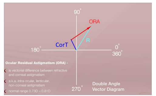
ORA is defined as the vectorial difference between the corneal topographical astigmatism and the refractive cylinder (R) – it is expressed in dioptres (D). Shown here is a double-angle vector diagram.
CorT = corneal topographic astigmatism; D = diopter; ORA = ocular residual astigmatism; R = refractive cylinder
The ORA parameter should be calculated routinely preoperatively for every patient seeking to have refractive laser surgery. This will determine how much of the astigmatism can be corrected with the surgery and allow the surgeon to advise the patient of potential outcomes. If the magnitude and orientation of the refractive and corneal astigmatism are identical, then the ORA will be zero and all the refractive and corneal astigmatism can potentially be corrected. It is important to note that the manifest refractive cylinder includes all the astigmatic components from the front of the cornea all the way back to the occipital cortex.
What can be done with ocular residual astigmatism?
If treatment parameters are based solely on refractive parameters, taking no account of the corneal astigmatism, then all the calculated ORA will be left on the cornea postoperatively. Conversely, if the treatment parameters are based entirely on the cornea (that is topography-guided), then all the ORA will remain in the refraction. There is no escaping any inherent difference in refractive/corneal astigmatism. Distributing the ORA to both the cornea and the refraction and including the corneal astigmatism parameters in the refractive treatment plan reduce the amount of corneal astigmatism remaining compared with the treatment based only on refractive parameters. This can be done using the method of vector planning. By placing emphasis on the ORA, optimal amounts of corneal and refractive astigmatism can be targeted postoperatively rather than leaving the entire amount of the ORA either on the cornea or in the refraction. Previous studies have shown an emphasis of 60% on the ORA towards refraction and 40% to cornea, and this is a good default point when treating astigmatism using vector planning.5–8 It is important to note that the total of the target corneal astigmatism and target refractive cylinder is always equal to the ORA regardless of the percentage of emphasis on the ORA. Any refractive cylinder targeted is offset by a spherical component that is half the magnitude of the cylinder to target a spherical equivalent of zero. This then is less perceived by the patient than the plano/cylinder left postoperatively in the refraction.
Studies on vector planning have shown a significant reduction in corneal astigmatism postoperatively compared with treatments using only refractive parameters.7,8 At the same time, the refractive outcomes are equivalent or better than those achieved using only 100% refractive treatment parameters. The benefit of maximally reducing corneal astigmatism is in maintaining the quality of vision – there is less possibility of ghosting, arcing, starbursts and haloes, known as GASH, due to high amounts of corneal astigmatism remaining postoperatively.9
Let us consider all this with an example. Figures 2 and 3 show an example where there is a significant preoperative ORA of 1.24D. By distributing some of this ORA to both the cornea and the refraction using vector planning, the maximum amount of astigmatism is treated, targeting 0.74D @ 96 on the cornea instead of the total ORA of 1.24D@96 as would be the case if treated using only refractive parameters. The total amount of ORA of 1.24D would be left in the refraction postoperatively if the treatment was planned based on 100% topography parameters. The other benefit of addressing the ORA using vector planning to note is that the corneal target orientation is now closer to with-the-rule astigmatism of 96° than preoperatively at an oblique 62°, which renders it more favourable (Figure 4).
Figure 2: Example of corneal astigmatism
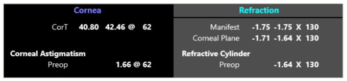
Both corneal and refractive parameters are used in the method of vector planning to treat astigmatism. In this example, the corneal topographical astigmatism measures 1.66D@62 and the refractive cylinder measures -1.64D×130 at the corneal plane.
CorT = corneal topographical astigmatism; Preop = preoperative.
Figure 3: The ocular residual astigmatism is calculated as 1.24D×6 (orange), and the emphasis on the ocular residual astigmatism is based on 60% refractive parameters (cyan) and 40% corneal parameters (violet) using vector planning
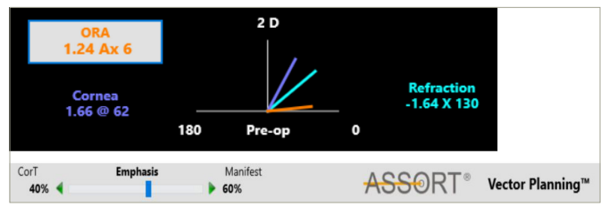
Ax = axis; CorT = corneal topographical astigmatism; D = diopter; Manifest = manifest refraction; ORA = ocular residual astigmatism; Pre-op = preoperative.
Figure 4: The targeted corneal and refractive parameters are 0.74D and 0.49D, respectively. Note the zero targeted spherical equivalent by offsetting the targeting refractive cylinder with a sphere of half the magnitude
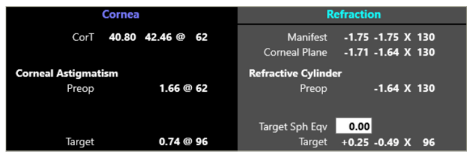
CorT = corneal topographic astigmatism; Eqv = Spherical Equivalent; Preop = preoperative; Sph = sphere
Figure 5 displays the orientation of the laser beam for the astigmatism treatment. With vector planning, the treatment is closer to the corneal astigmatism orientation than if treated using only manifest refraction. The treatment plan incorporated into the laser devices using vector planning is significantly different from that based only on refractive parameters (Figure 6).
Figure 5: Orientation of the laser beam for astigmatism treatment
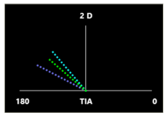
With vector planning, the astigmatic laser treatment lies between the refractive (cyan) and corneal (violet) parameter orientation as shown in green.
D = diopters; TIA = target induced astigmatism vector.
Figure 6: Vector planning
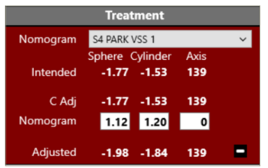
The vector planning treatment input into the laser device (‘Adjusted’ line in the diagram) is different from the manifest refraction of -1.71DS/-1.64DC × 130. Note that the diagram also shows a spherical and cylindrical nomogram adjustment.
C Adj = cylinder adjustment; DC = dioptre cylinder; DS = dioptre sphere; S4 PARK VSS 1 = nomogram name for ViSX VSS excimer laser
Which measure of corneal astigmatism should be included in the vector planning method to optimally treat astigmatism?
The corneal topographic astigmatism parameter (CorT anterior) was first reported in a study published in the Journal of Cataract and Refractive Surgery in 2012, showing that a more accurate measure of corneal astigmatism could be obtained using the data from all the Placido rings in topography than using the data from a single Placido ring as is the case with simulated keratometry values.10 This study was further evaluated and expanded on in the Journal of Refractive Surgery in 2015 to include the total corneal power using the data from both the anterior and posterior corneal surfaces.11 Known as the CorT Total, it was shown that this parameter was more accurate in measuring the total corneal power than CorT anterior. This CorT Total parameter is, therefore, recommended to be used when planning treatments with vector planning. Other measures of total corneal astigmatism can be included when planning using vector planning, and the ORA can be calculated for all corneal measures versus refractive cylinder to establish which is the most accurate – this will be reflected by the smallest ORA magnitude.
Dividing the cornea into two (hemidivisional treatment)
An approach considering further customization of the treatment of astigmatism involves conceptually dividing the cornea into two halves. In this way, the method of vector planning can be further refined and used to treat each half of the cornea with different emphases on the refraction and corneal parameters. The planned emphases will depend on the amount of total corneal astigmatism in each half. At this point in time, refractive laser devices treat the cornea with one overall spherocylindrical treatment predominantly as measured by manifest refraction. If corneal parameters are included in the treatment plan, such as vector planning, then these are based on one measure of total corneal astigmatism. With advances in tomography, multiple measures of total corneal astigmatism can now be evaluated, so that each half of the cornea can have its own measure of total corneal astigmatism, and the one common manifest refractive cylinder can be used in combination with vector planning to further customize the treatment of astigmatism.
The method of treating astigmatism using vector planning for the two halves of the cornea has recently been published.1,12 To delineate the two halves of the cornea, the flat meridian of the CorT Total is recommended. This then allows for a total corneal measure for each half of the cornea. Therefore, the vector planning method uses the total corneal power for one half of the cornea together with the common manifest refractive cylinder for both halves of the cornea. The emphasis on the ORA for each half is selected by the surgeon to target the distribution of the ORA to both the cornea and refraction postoperatively. The two halves can have different emphases on the ORA depending on the mismatch between corneal and refractive astigmatism parameters. If, for example, there is much more corneal astigmatism than a refractive cylinder in one half of the cornea, then there would be more emphasis placed on reducing the corneal astigmatism and vice versa. In Figure 7, preoperatively, there is more corneal astigmatism in the inferior half than the superior (shown in purple), and hence, the emphasis on the ORA is more towards treating corneal astigmatism than the superior half (40% corneal emphasis superiorly compared with 50% emphasis inferiorly). In theory, there are two steps involved: (1) maximally reducing astigmatism for each half of the cornea and (2) regularizing any remaining corneal astigmatism in a symmetric, orthogonal bow-tie appearance on tomography, potentially improving best corrected visual acuity. In practice, this is all done in one treatment – reducing and regularizing astigmatism as well as correcting any spherical error. Currently, we cannot avail this astigmatism treatment using laser devices, but the software for the treatment parameters is freely available online at both www.assort.com and www.isrs.org.13,14 This concept should stimulate interest among laser manufacturers to avail such customized treatments to doctors for use with LASIK (laser assisted in situ keratomileusis), PRK (photorefractive keratectomy) and lenticule extraction procedures.
Figure 7: Hemidivisional reduction and regularization
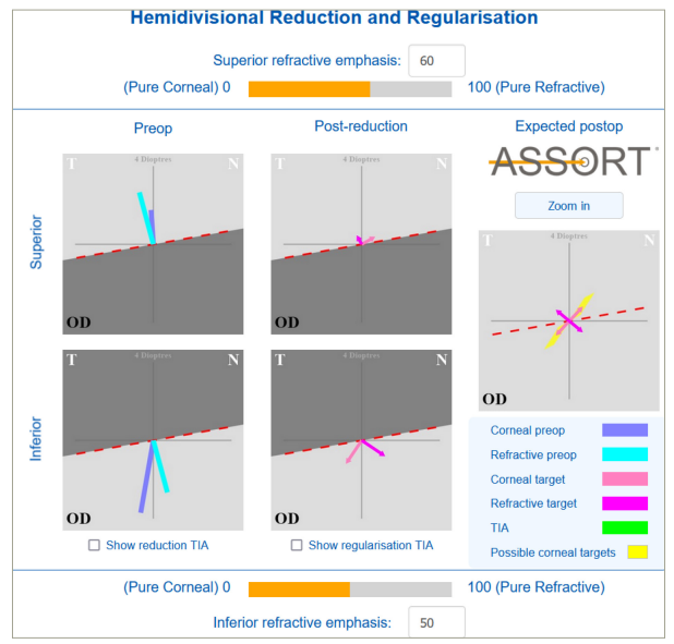
A theoretical approach to treating regular and irregular astigmatism by conceptually dividing the cornea into two halves.
N = nasal; OD = right eye; Preop = preoperative; Postop = postoperative; T = temporal; TIA = target induced astigmatism vector
The calculation of ORA preoperatively and the method of vector planning are being used routinely across the world to treat astigmatism. Data from the free calculators available on the ASSORT® website shows that over a 6-month period, more than 22 countries are using the ORA calculator and more than 1,500 calculations are being done.13 Regarding the vector planning calculator, there are more than 19 countries using the method and 1,900 calculations are being done. These numbers show the increasing adoption of the importance of vector planning and the ability to optimally treat astigmatism. The next step will require laser manufacturers to adopt the hemidivision technology to aid surgeons in further enhancing astigmatism outcomes.1,12
Furthermore, a recent article in the Journal of Refractive Surgery which focuses on the most cited articles in refractive surgery spanning over 40 years, ranked Alpins’ 1993 article discussing the variance between spectacle and corneal astigmatism as the 14th foundational paper with 354 citations (now 444). Additionally, the Alpins’ method in the 2001 article holds the 18th position overall with 361 citations (now 471).2,15,16
With vector planning gaining traction in its adoption worldwide to most effectively treat astigmatism using LASIK, PRK or lenticule extraction procedures, laser manufacturers need to take note of the next level in the treatment of regular and irregular astigmatism and make it possible for the surgeon to further customize the treatment of astigmatism to maximize success by treating different parts of the cornea using specific combined refractive and corneal measures.


