touchOPHTHALMOLOGY touchOPHTHALMOLOGY
Oculoplastic Surgery
Read Time: 3 mins

Trichofolliculoma of the Palpebral Conjunctiva: A Case Report

Copy Link
Published Online: May 26th 2021 touchREVIEWS in Ophthalmology. 2021;15(1):38–9. DOI: https://doi.org/10.17925/OPHT.2021.15.1.38
Authors: Spencer C Cleland, Christopher P Schiefer, Paige J Richards, Jaclyn Gurwin, Heather AD Potter, Cat N Burkat
Quick Links:
Abstract
Article
Article Information
Abstract:
Overview

Background: Trichofolliculoma is an uncommon, benign cutaneous neoplasm with hair follicle differentiation, which typically affects the head and neck. However, there are no reported cases of a trichofolliculoma emanating from the palpebral conjunctiva. Case presentation: This case report describes a 15-year-old male who presented following several years of intermittent foreign body sensation of the left eye. Double eversion of the upper eyelid demonstrated numerous long dark hairs originating from a pink nodule in the superolateral fornix. Excisional biopsy with histopathologic analysis demonstrated a dilated hair follicle surrounded by secondary and tertiary hair follicles, which is consistent with the features of a trichofolliculoma. Conclusions: Trichofolliculoma can be seen on the palpebral conjunctiva. Complete surgical excision is recommended to reduce the rate of recurrence and scar formation.

Keywords

Trichofolliculoma, conjunctiva, ocular pathology, dermatopathology, case report

Article:

Trichofolliculoma is a rare, benign skin tumour arising from a hair follicle containing a large central cystic hair follicle with branching epithelial strands and buds.1–5 Trichofolliculomas typically affect the head and neck, appear flesh-coloured, dome-shaped, and may also have a tuft of hair emanating from a central pit.1–5 We report a case of a 15-year-old male with a lesion of the palpebral conjunctiva with clinical and histopathologic features consistent with trichofolliculoma.

Case presentation

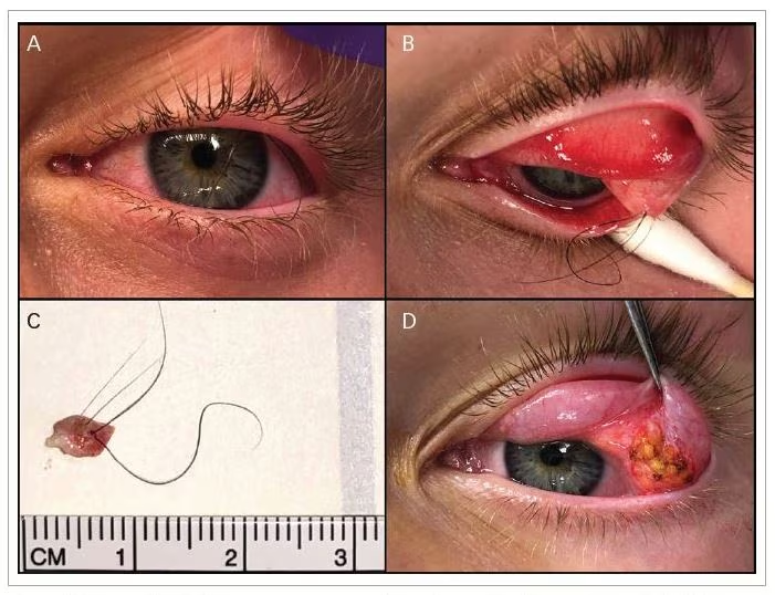
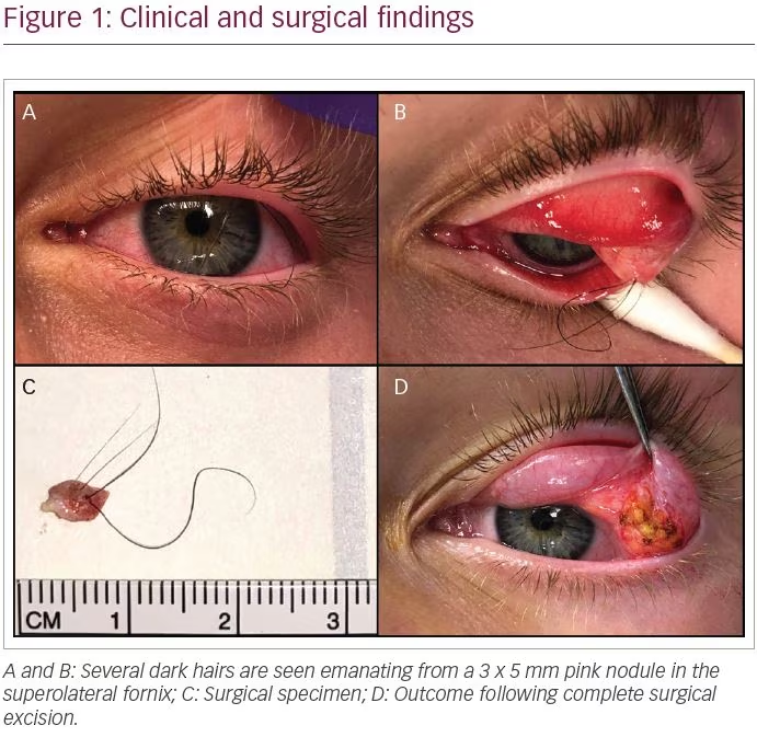
A healthy 15-year-old boy with several years of intermittent foreign-body sensation in the left eye presented with worsening eye irritation. A slit lamp examination revealed several long dark hairs emanating from the superior fornix and traversing the surface of the cornea (Figure 1A). Double eversion of the left upper eyelid with gentle pressure on the globe revealed that these hairs were originating from an elevated pink nodule of the palpebral conjunctiva in the superolateral fornix (Figure 1B). In an attempt to relieve the irritation, several hairs were removed at the slit lamp. However, the procedure was tolerated poorly, and the patient subsequently underwent complete surgical excision of the lesion under sedation. The nodule was found to be adherent to, but separate, from the lacrimal gland (Figure 1C and D). Histologic analysis demonstrated a dilated hair follicle containing several hair fragments surrounded by numerous secondary and tertiary hair follicles, consistent with trichofolliculoma (Figure 2). The postoperative course was uncomplicated, with complete resolution of the foreign body sensation.

Discussion and conclusion

Trichofolliculoma is an uncommon, benign cutaneous neoplasm with hair follicle differentiation, which typically affects the head and neck.1–5 These hamartomas can affect men and women of all ages, and represent 1.3% of all eyelid adnexal tumors.1,2 Clinically, trichofolliculomas are small, smooth, pearly nodules, and often contain a central pore with wisps of immature hair.1–5 This case shared all of these features; however, this patient’s lesion was unique due to its location in the superolateral conjunctival fornix adjacent to the lacrimal gland. Trichofolliculomas are typically seen cutaneously, given their hair follicle origin, so it is unusual to observe the lesion on a mucous membrane. Although trichofolliculoma is well known to originate on the skin of the eyelids,5 this is the first reported case of trichofolliculoma arising from the palpebral conjunctiva reported in the literature.

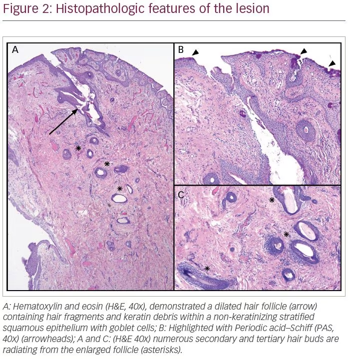
The characteristic histologic findings of a trichofolliculoma include a dilated follicle within the upper dermis containing keratin debris and hair shafts.1–5 In addition, numerous smaller hair follicles radiate from the primary central pore.1–5 These features are evident in this case (Figure 2). However, there is also non-keratinizing stratified squamous epithelium with goblet cells, highlighted by Periodic acid–Schiff stain (Figure 2B), consistent with the trichofolliculoma arising from the palpebral conjunctiva. Given the presence of a central pore and cystic space continuous with the surface, the differential diagnosis also included dilated pore of Winer and pilar sheath acanthoma.4,6 However, this lesion lacks elongated rete ridges or lobules of infundibular cells extending from the enlarged follicle, which are seen in dilated pore of Winer and pilar sheath acanthoma, respectively.4,6,7. Dermoid cyst was also considered due to the lesion’s location in the superolateral fornix, though the absence of other adnexal structures, such as sebaceous glands, contiguous with the lesion made this diagnosis less likely.

The treatment of choice for a trichofolliculoma is complete surgical excision.1,8 Despite being a benign tumour, incomplete excision of a trichofolliculoma has been associated with recurrence.1,5,8 Also, merely epilating the lesion can cause an inflammatory reaction and subsequent scar tissue formation.1 Fortunately, this patient achieved a successful postoperative outcome with no injury to the lacrimal gland.

Article Information:
Disclosure

Spencer C Cleland, Christopher P Schiefer, Paige J Richards, Jaclyn Gurwin, Heather AD Potter and Cat N Burkat have no financial or non-financial relationships or activities to declare in relation to this article.

Compliance With Ethics

All procedures were followed in accordance with the responsible committee on human experimentation and with the Helsinki Declaration of 1975 and subsequent revisions, and informed consent was received from the patient involved in this case study.

Review Process

Double-blind peer review.

Authorship

The named authors meet the International Committee of Medical Journal Editors (ICMJE) criteria for authorship of this manuscript, take responsibility for the integrity of the work as a whole, and have given final approval for the version to be published.

Correspondence

Spencer C Cleland, University of Wisconsin, Department of Ophthalmology and Visual Sciences, 600 Highland Avenue, K6/448 Clinical Science Center, Madison, WI 53792, USA. E: scleland0331@gmail.com

Support

No funding was received for the publication of this article.

Access

This article is freely accessible at touchOPHTHALMOLOGY.com © Touch Medical Media 2021

Received

2021-01-14

References

  1. Kapoor AG, Vijitha VS, Mittal R. Trichofolliculoma of the eyelid margin.Ophthal Plast Reconstr Surg. 2019;35:e74–6.
  2. Ozdal PC, Callejo SA, Codere F, Burnier MNJ. Benign ocular adnexal tumours of apocrine, eccrine or hair follicle origin. Can J Ophthalmol. 2003;38:357–63.
  3. Peterdy GA, Huettner PC, Rajaram V, Lind AC. Trichofolliculoma of the vulva associated with vulvar intraepithelial neoplasia: Report of three cases and review of the literature. Int J Gynecol Pathol.2002;21:224–30.
  4. Tellechea O, Reis JP, Gameiro AR, et al. Benign follicular tumors. An Bras Dermatol. 2006;90:780–98.
  5. Phan K, Goyal S, Ahn E, et al. Trichofolliculoma of the eyelid: systematic review. Biomed Dermatology.2020;4:3.
  6. Klein W, Chan E, Seykora JT. Tumors of The Epidermal Appendages. In: Elder DE, Elenitsas R, Johnson Jr BL, Murphy GF, eds, Lever’s Histopathology of the Skin. Philadelphia, PA: Lippincott Williams & Wilkins; 2005.
  7. Jakobiec FA, Bhat P, Sutula F. Winer’s dilated pore of the eyelid. Ophthal Plast Reconstr Surg. 2009;25:411–3.
  8. Morton AD, Nelson CC, Headington JT, Elner VM. Recurrent trichofolliculoma of the upper eyelid margin. Ophthal Plast Reconstr Surg. 1997;13:287–8.

Further Resources

Share this Article
Related Content In Oculoplastic Surgery
  • Copied to clipboard!
    accredited arrow-down-editablearrow-downarrow_leftarrow-right-bluearrow-right-dark-bluearrow-right-greenarrow-right-greyarrow-right-orangearrow-right-whitearrow-right-bluearrow-up-orangeavatarcalendarchevron-down consultant-pathologist-nurseconsultant-pathologistcrosscrossdownloademailexclaimationfeedbackfiltergraph-arrowinterviewslinkmdt_iconmenumore_dots nurse-consultantpadlock patient-advocate-pathologistpatient-consultantpatientperson pharmacist-nurseplay_buttonplay-colour-tmcplay-colourAsset 1podcastprinter scenerysearch share single-doctor social_facebooksocial_googleplussocial_instagramsocial_linkedin_altsocial_linkedin_altsocial_pinterestlogo-twitter-glyph-32social_youtubeshape-star (1)tick-bluetick-orangetick-red tick-whiteticktimetranscriptup-arrowwebinar Sponsored Department Location NEW TMM Corporate Services Icons-07NEW TMM Corporate Services Icons-08NEW TMM Corporate Services Icons-09NEW TMM Corporate Services Icons-10NEW TMM Corporate Services Icons-11NEW TMM Corporate Services Icons-12Salary £ TMM-Corp-Site-Icons-01TMM-Corp-Site-Icons-02TMM-Corp-Site-Icons-03TMM-Corp-Site-Icons-04TMM-Corp-Site-Icons-05TMM-Corp-Site-Icons-06TMM-Corp-Site-Icons-07TMM-Corp-Site-Icons-08TMM-Corp-Site-Icons-09TMM-Corp-Site-Icons-10TMM-Corp-Site-Icons-11TMM-Corp-Site-Icons-12TMM-Corp-Site-Icons-13TMM-Corp-Site-Icons-14TMM-Corp-Site-Icons-15TMM-Corp-Site-Icons-16TMM-Corp-Site-Icons-17TMM-Corp-Site-Icons-18TMM-Corp-Site-Icons-19TMM-Corp-Site-Icons-20TMM-Corp-Site-Icons-21TMM-Corp-Site-Icons-22TMM-Corp-Site-Icons-23TMM-Corp-Site-Icons-24TMM-Corp-Site-Icons-25TMM-Corp-Site-Icons-26TMM-Corp-Site-Icons-27TMM-Corp-Site-Icons-28TMM-Corp-Site-Icons-29TMM-Corp-Site-Icons-30TMM-Corp-Site-Icons-31TMM-Corp-Site-Icons-32TMM-Corp-Site-Icons-33TMM-Corp-Site-Icons-34TMM-Corp-Site-Icons-35TMM-Corp-Site-Icons-36TMM-Corp-Site-Icons-37TMM-Corp-Site-Icons-38TMM-Corp-Site-Icons-39TMM-Corp-Site-Icons-40TMM-Corp-Site-Icons-41TMM-Corp-Site-Icons-42TMM-Corp-Site-Icons-43TMM-Corp-Site-Icons-44TMM-Corp-Site-Icons-45TMM-Corp-Site-Icons-46TMM-Corp-Site-Icons-47TMM-Corp-Site-Icons-48TMM-Corp-Site-Icons-49TMM-Corp-Site-Icons-50TMM-Corp-Site-Icons-51TMM-Corp-Site-Icons-52TMM-Corp-Site-Icons-53TMM-Corp-Site-Icons-54TMM-Corp-Site-Icons-55TMM-Corp-Site-Icons-56TMM-Corp-Site-Icons-57TMM-Corp-Site-Icons-58TMM-Corp-Site-Icons-59TMM-Corp-Site-Icons-60TMM-Corp-Site-Icons-61TMM-Corp-Site-Icons-62TMM-Corp-Site-Icons-63TMM-Corp-Site-Icons-64TMM-Corp-Site-Icons-65TMM-Corp-Site-Icons-66TMM-Corp-Site-Icons-67TMM-Corp-Site-Icons-68TMM-Corp-Site-Icons-69TMM-Corp-Site-Icons-70TMM-Corp-Site-Icons-71TMM-Corp-Site-Icons-72