Until recently, it seemed inconceivable to most vision researchers and ophthalmologists alike that there could be an unrecognised class of photoreceptor within the eye. After all, the eye was the best understood part of the central nervous system. One hundred and fifty years of intensive research had explained how we see: photons are detected by the rods and cones and their graded potentials are assembled into a crude image by the inner retina, followed by advanced visual processing in the brain. This depiction of vision left no room for an additional class of photoreceptor.
Until recently, it seemed inconceivable to most vision researchers and ophthalmologists alike that there could be an unrecognised class of photoreceptor within the eye. After all, the eye was the best understood part of the central nervous system. One hundred and fifty years of intensive research had explained how we see: photons are detected by the rods and cones and their graded potentials are assembled into a crude image by the inner retina, followed by advanced visual processing in the brain. This depiction of vision left no room for an additional class of photoreceptor.
However, this conventional view of retinal organisation has now been overturned. We now appreciate that the rods and cones are not the only photosensory neurons of the eye. This discovery has its origins in attempts to understand how endogenous 24-hour body clocks (circadian clocks) are regulated by light, and this is where this article will start. However, this third class of ocular photoreceptor does much more than regulate the body clock, and its contribution to a range of light detection tasks should now be factored into all assessments of clinical blindness.
Light gives both a spatial and a temporal dimension to our world. Most organisms possess an endogenous 24-hour circadian timing system that ‘fine-tunes’ physiology and behaviour to the varying demands of the day–night cycle. However, such a temporal programme is useful only if biological time remains synchronised to the solar day. The behavioural and physiological disruption we experience during ‘jet-lag’ illustrates the importance of the synchronised circadian system.
Most organisms, including humans, have evolved to use the dawn/dusk light transition as the main zeitgeber (time-giver) to adjust circadian time to local time, a process termed photoentrainment. In mammals, the master circadian pacemaker is located within small paired nuclei of the anterior hypothalamus called the suprachiasmatic nuclei (SCN), and receives a direct retinal projection via the retinohypothalamic tract. Eye loss in mammals blocks photoentrainment. Therefore, mammalian eyes perform two quite different sensory tasks: their familiar function is to collect and process light to generate an image of the world, while their less wellrecognised role is to provide measures of environmental irradiance over the period of dawn and dusk to facilitate photoentrainment. Such divergent responses to light were difficult to reconcile within the known physiology of the rods and cones, which integrate photons over extremely short time periods.1
In the early 1990s, mice homozygous for gene defects, e.g. retinal degeneration (rd), and lacking any visual responses to light were examined to determine the impact of rod/cone loss on photoentrainment. Remarkably, rd/rd mice lacking functional rods and most cones showed normal circadian responses to light.2 These and a host of subsequent experiments, including studies in humans with genetic defects of the eye,3,4 showed that the processing of light information by the circadian and classic visual systems must be different, and raised the possibility that the eye may contain an additional non-rod, noncone photoreceptor. This was a supposition that was greeted with derision by referees and funding bodies alike, based largely on the assumption that only a small number of rods and/or cones were necessary for normal photoentrainment of the clock. To test this assumption, a mouse was engineered in which all rods and cones were ablated (rd/rd cl). Such genetic lesions had little effect on circadian responses to light, although loss of the eyes completely abolished this capacity.5,6 The rd/rd cl mouse model also proved invaluable in showing that a range of other irradiance detection tasks do not require the rods and cones.
In mammals, light-induced pupil constriction is regulated by the rods and cones, yet multiple studies have shown that pupil constriction still occurs after profound damage to these photoreceptors. Not unreasonably, it was assumed that the residual light-induced pupil constriction was due to the survival of a few intact rod and/or cone photoreceptors and/or retinal re-wiring.7 The rd/rd cl mouse allowed an explicit test of this assumption. Pupil measurements were undertaken in rd/rd cl mice and showed that these animals were fully able to constrict their pupils under bright light conditions.8
It is important to stress that, unlike in circadian responses to light, rod and cone photoreceptor loss has a clear impact on the sensitivity of pupil constriction. It appears that the rods and cones mediate constriction under relatively dim and transitory responses to light, while the novel receptors drive constriction under sustained bright light conditions.
By 2001, it was clear that novel photoreceptors exist within the eye and that they regulate a variety of responses to environmental brightness; however, their cellular identity remained unclear. Retinal ganglion cells (RGCs) had been implicated as these photoreceptors,9 but the final proof came from two independent approaches: one in rats, the other in rd/rd cl mice. Both showed that the retina contains a small number of photosensitive RGCs (pRGCs). In rats, a subpopulation of RGCs was labelled by retrograde dye injected into the SCN. The retina was removed and the electrical activity of individually labelled RGCs was monitored by intracellular recording. These cells responded to bright light. This in itself was no surprise because the rods and cones were present in these retinae. However, light-evoked depolarisations persisted in the presence of a cocktail of drugs considered sufficient to block all retinal intercellular communication, and even persisted in RGCs that were micro-dissected from the surrounding retinal tissue.10
The second approach exploited the rd/rd cl mouse retina in combination with calcium (Ca2+) imaging. This technique is capable of detecting small changes in the concentration of intracellular Ca2+ in large numbers of individual living cells across the entire retina. The use of the rd/rd cl mouse meant that pharmacological or surgical isolation of ganglion cells from the rod and cone photoreceptors was unnecessary. This study demonstrated that approximately 3% of the neurons in the RGC layer responded to light, but after blocking gap junctions the number of RGCs responding to light dropped to ~1%. These results showed that there exists an extensive network of photosensitive RGCs that can be uncoupled by application of gap-junctional blockers. Furthermore, three types of light-evoked Ca2+ influx were observed in these neurons: a sustained response, a transient response and a repetitive response. Collectively, the studies in rd/rd cl mice identified a heterogeneous coupled syncytium of pRGCs.11
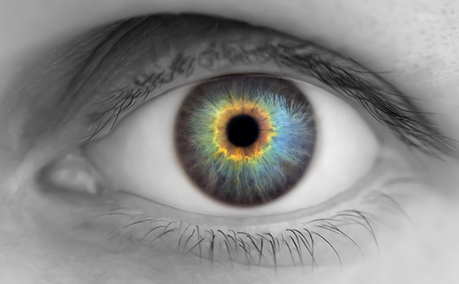
The photopigment of the pRGCs was defined in the first instance by action spectroscopy. This powerful approach rests on the fact that a photopigment has a characteristic absorbance spectrum or profile that describes the likelihood of photons being absorbed at different wavelengths. Thus, a description of the spectral sensitivity profile (action spectrum) of any light-dependent response will describe the absorbance spectrum of the photopigment on which the response is based. All vitamin-A-based photopigments have a characteristic absorption spectrum. This means that although the peak sensitivity of the pigment may vary widely across the visible spectrum (ultraviolet at 360nm to deep red at 750nm), all of these pigments have the same basic shape.12 The first full-action spectrum to define the nature of the photopigment of the pRGCs studied pupil constriction in rd/rd cl mice. The results described a previously undefined opsin/vitamin-A-based photopigment with peak sensitivity in the ‘blue’ (λmax ~479nm) region of the spectrum, which was tentatively designated opsin photopigment/ λmax 479nm (OP479).8
Since 2001, a plethora of action spectra from mice to man have been deduced for a range of irradiance responses to light. These include the light responses of pRGCs in mice,13 rats10 and primates,14 and span pupil constriction, phase-shifting circadian rhythms and plasma melatonin suppression, together with irradiance-dependent regulation of human retinal-cone function.15 All of these action spectra point to the existence of a coherent single novel opsin photopigment (OP479) with a λmax of around 480nm. It remains unclear what advantage a λmax of around 480nm may confer on such diverse species. One possibility is that the pRGCs are tuned to the dominant wavelength of light at twilight. During twilight the sun is close to the horizon and there is relative enrichment of ‘blue’ light in the dome of the sky because of the preferential scattering of short wavelengths of light passing obliquely through the atmosphere.
Although the photopigment of the pRGCs had been defined using action spectroscopy, the opsin gene of OP479 remained unknown – but not for long. The melanopsin gene family, also designated Opn4, was first identified in Xenopus photosensitive pigment cells, i.e. melanophores (hence the name melanopsin),16 and then orthologues were isolated from other vertebrate classes, including zebrafish17 and several mammalian species, including humans and mice.18 Melanopsin was immediately implicated as the photopigment, as it is expressed in pRGCs and ablation of the melanopsin gene abolishes the pRGC responses to light.10,11 Furthermore, mice in which rods, cones and melanopsin have all been ablated fail to show circadian or pupil responses to light, suggesting that these three classes of photoreceptor can fully account for all light detection within the eye.13 Although highly suggestive, the melanopsin knockout data could not demonstrate that melanopsin was the photopigment of the pRGCs. Gene ablation alone can only indicate that a gene is important; biochemistry on the protein product is usually required to define its function. This critical matter was finally addressed by three groups using heterologous expression of either human or murine melanopsin in neuro2A cells,19 human embryonic kidney (HEK)-293 cells20 and Xenopus oocytes.21 In each expression system, melanopsin expression was fully sufficient to drive a retinal-dependent light cascade in vitro. Intriguingly, several recent studies have also shown that melanopsin may trigger a signalling cascade that more closely resembles the signalling pathway of an invertebrate photopigment rather than a rod or cone.22–25
Thus, the mammalian retina contains a population of melanopsin-based pRGCs that are used to measure environmental irradiance and modulate diverse physiological responses to light, including circadian physiology and pupil constriction. The initial assumption was that there would be little functional overlap between pRGCs and the rods/cones. However, we now know that there is complex cross-talk between these image- and non-image-forming ocular photoreceptors. This is obvious in the case of the pupil light reflex, where the loss of the rods and cones leads to a marked decrease in the sensitivity of the response. However, in the case of the circadian system the role of the rods and cones is far less clear. The loss of rods and cones in rd/rd cl mice results in only subtle effects on circadian entrainment and, as may be predicted, the loss of rods, cones and pRGCs abolishes all light responses.13
However, the loss of melanopsin in the pRGCs alone does not abolish circadian photosensitivity, but rather attenuates circadian responses to light. It seems that under these circumstances the rods and/or cones can partially compensate for pRGC loss of function. Additional evidence for rod, cone and pRGC interaction comes from studies on the macaque. Intracellular recording from melanopsin pRGCs have shown that the S-wavelength cones (λmax ~435nm) attenuate the light responses of pRGCs, while the inputs from the rods, medium (M) (λmax ~530nm) and long (L) (λmax ~60nm) wavelength cones provide an excitatory input.14 The explanation for this opponent interaction remains unclear.22
Research on the pRGCs has been largely confined to animal models, and the extent to which the findings in rodents and non-human primates are mirrored in humans has been the subject of much debate. The participation of two profoundly blind subjects lacking functional rods and cones has allowed an exploration of this question. The first finding was that human subjects lacking rods and cones are still capable of regulating their circadian physiology and behaviour.26 On the basis of these findings, and by analogy to the studies in rodents,8 it was reasoned that some pupil reactivity to bright light should also be retained, despite the clinical reports that subjects are unresponsive to the brief light exposure from a pen torch.
One individual was examined in detail and clearly possessed a functioning pupillomotor system responsive to bright light. Furthermore, the pupil constriction response was spectrally tuned, peaking (λmax) around 480nm, which corresponds well to the action spectra for pRGCs in both human (483nm)15 and non-human primates (482nm),14 but not the λmax of human rods (~498nm) or S-, M- and Lwavelength cones (λmax ~420, 534 and 563nm, respectively).
Humans appear to possess a pRGC system that to a high degree resembles that of rodents and non-human primates. Furthermore, the demonstration of a pupil response in a rodless/coneless individual raises important issues regarding the significance of this assay in defining blindness. Currently, unreactive pupils in response to brief torch pen examination are considered clinically to be a sine qua non of profound blindness of retinal origin.26 Ideally, such an examination should involve exposure to bright light over many seconds.
What else may the pRGCs regulate in humans? Recent findings in primates showed that the pRGCs project to the dorsal lateral geniculate nucleus (dLGN), which is the thalamic relay that provides a direct input to the visual cortex.14 This raised the possibility that these photoreceptors may contribute to an individual’s ability to detect or even experience some awareness of light, triggering either a conscious response or, in the absence of a percept, a response analogous to blindsight – defined as an above chance ability to detect the presence of a light stimulus in the absence of a conscious percept. Therefore, a rodless/coneless subject was examined to determine whether a given light stimulus could be detected when presented in the first or second of two temporal intervals in a two-alternative forced-choice paradigm (2AFC). After some initial hesitancy on being asked to report the presence of visual stimuli of which she was nominally unaware, the subject was able to correctly identify the interval in which a 481nm test light (at threshold intensity) appeared, but failed to detect light at longer or shorter wavelengths (420, 460, 500, 515, 540, 560 and 580nm).
Collectively, these results provide strong evidence that pRGCs can, at some level, contribute to our awareness of environmental light. These surprising findings suggest a greater functional overlap of the pRGCs and rod/cone subsystems than was previously assumed.26
The discovery of a third photoreceptor system within the eye based on melanopsin pRGCs argues that the clinical diagnosis of ‘complete’ blindness should assess the state of both the rod/cone and pRGC photoreceptive systems. We now appreciate that eye loss plunges individuals into a world that lacks both vision and a proper sense of time, and clinical guidelines should incorporate this information. If a ‘blind’ individual shows a bright-light-dependent pupil constriction, he or she should be encouraged to expose his or her eyes to sufficient daytime light to maintain normal circadian entrainment and sleep–wake timing.
Furthermore, patients with diseases of the inner retina that result in RGC death (e.g. glaucoma) are at particular risk of circadian rhythm and sleep disruption. Such individuals should receive counselling regarding the problems of sleep disruption27 and would be strong candidates for treatment with appropriately timed melatonin, which has been shown to consolidate sleep timing in patients suffering eye loss.28,29