Optical lens filtration therapy began with FL-41 lenses that were developed in the late 1980s and first reported in 1991 by Wilkins and Wilkinson.1 They were originally designed to improve workplace productivity by reducing eye strain and headaches induced by fluorescent lighting. They were designed to allow only 10% light filtration from 400–550 nm with a gradual increase in transmission from there with increased wavelength.1 The resulting lenses offered peak filtration at 480 nm and showed initial anecdotal efficacy in patients with agoraphobia, photosensitive epilepsy, post-traumatic photosensitivity, and eye strain.1 A subsequent study by Good et al., in the same year, showed a reduction in migraine frequency in children wearing the lenses.2 This initial design occurred many years before the discovery of intrinsically photosensitive retinal ganglion cells (IPRGCs) and its relation to the pathophysiology of photosensitivity.3,4 This article will discuss the mechanism and efficacy for various applications of optical lens filters and wavelength-specific blockade.
The literature was reviewed with PubMed queries for “FL-41”, “lens filters photosensitivity”, “lens filters photophobia”, “axon optics photophobia”, and “lens filters melanopsin”. Articles were identified from the year 1990 to the present that discussed the pathophysiology of photophobia and optical filtration therapy. References from included articles were also examined in search of other pertinent articles.
Mechanism
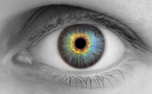
Blue-blocking lenses, such as the original FL-41, preferentially block shorter wavelengths of light in the visible spectrum, usually including significant filtration around 480 nm.1,5 This appears to be a particularly important wavelength, because it is also the wavelength of light that induces phototransduction in IPRGCs, which were discovered around a decade later.3,4 These retinal ganglion cells are labeled “intrinsically photosensitive” because they are able to transduce light signals independent of traditional rod or cone photoreceptors.3,6–10 They have been shown to have involvement in both circadian rhythms and the pupillary light reflex.3,4 Modulation of IPRGC phototransduction is thought to be influenced by melanopsin, a biphasic pigment found in a IPRGCs that isomerizes between its all-trans and 11-cis forms upon exposure to 481 nm and 587 nm light, respectively.7 This means that the original blue-blocking lenses were unknowingly developed with a peak filtration that minimized the isomerization of melanopsin, and therefore, reduced rod–cone independent phototransduction by IPRGCs.
Multiple pathways have been theorized to mediate photophobia (e.g., trigeminal, sympathetic mediation),6,10,11 but one of those is directly mediated by IPRGCs, with signal transduction to nociceptive centers in the thalamus,6,8 an area that has shown positron emission tomography activation in photophobic patients with essential blepharospasm.12 This concurs with pre-existing evidence from Stringham et al. that shows that the symptoms of photophobia may increase with exposure to shorter wavelengths of light,13 and newer evidence from Zivcevska et al. that also showed greater sensitivity to light that corresponded to melanopsin’s peak spectral sensitivity in both healthy and clinical patients.14 The study by Stringham et al. was in three healthy subjects, and though it showed increased photosensitivity with shorter wavelengths, their action spectrum peaked around 520 nm, later than the melanopsin absorption spectrum.13 Additionally, a recent study by Marek et al. showed alleviation of blue-light (shorter wavelength) mediated photosensitivity in mice through application of a melanopsin antagonist.9
Therefore, theoretically, lenses that reduce exposure to light around 481 nm would decrease isomerization of melanopsin to its all-trans form and subsequent IPRGC-mediated phototransduction to nociceptive centers in the posterior thalamus. This is one proposed mechanism for the use of blue-blocking lenses as an adjunct treatment to a variety of conditions that champion photosensitivity as a prominent symptom. Other proposed mechanisms include increased comfort due to a reduction in overall light, sensitivity to a wavelength other than 480 nm, or the presence of a placebo effect when wearing tinted lenses.
Applications
Historical applications of tinted lenses have included treatment of patients with migraines, traumatic brain injury, benign essential blepharospasm (BEB), sleep difficulties, post-traumatic photosensitivity, and retinal dystrophies.2,5,9,15–23 In theory, however, they could be therapeutic in any patient with a photophobia-inducing condition, though they may be more effective in conditions whose nociceptive component relies more heavily on an IPRGC-mediated neural pathway. Various pathologies that may induce photophobia can be ocular (e.g., dry eye, uveitis, cone dystrophy), neurologic (e.g., optic neuritis, demyelinating disorders, migraine), or medication-related (e.g., lithium, benzodiazepines, chloroquine) in origin, among many other ailments (e.g., intracranial neoplasm, zinc deficiency).6,10 Additionally, the notion that optical filters may have clinical potential in the treatment of photophobia is supported by many patients experiencing photophobic symptoms after cataract surgery. The appearance of photophobic symptoms after the removal of a patient’s intrinsic light filter may suggest that a filter could be added to mitigate these same symptoms.
Migraine
Migraines were one of the first diseases targeted by optic-lens tinting due to their prominent photophobia. In 1991, Good et al. randomized 20 children with clinically diagnosed migraines to one cohort wearing blue-tinted and another wearing rose-tinted glasses. Only the patients wearing rose-tinted glasses (blue-light blocking) reported symptom reduction.2 A few years later, Main et al. reported that patients had a lower discomfort threshold to low wavelength light between migraine attacks (compared with medium and high wavelength).16 This has been reinforced with functional magnetic resonance imaging (fMRI) data that showed decreased cortical activation and subjective decreases in illusions and distortions in patients with migraine who wore precision optical tints when exposed to stressful striped images.24 To more precisely test this hypothesis, Hoggan et al. produced an optical notch filter (named for the shape of its light transmission curve) that more specifically blocks 480 nm light. This serves the same purpose as the traditional FL-41 tint, but it uses thin film technology instead of directly dying the lens to provide a more precise spectral filter. It was tested in patients with migraine, and symptoms were relieved with both a 480 nm and 620 nm filter.22 This further suggests that wavelength-specific blockade is likely a viable treatment option for patients migraine; however, it also presents the possibility that the improved comfort could be due to an overall reduction of light. The efficacy seen with 620 nm blockade is hypothesized to relate to the bi-stable nature of melanopsin and merits further investigation.22
Traumatic brain injury
Optical tinting has also shown promise in the treatment of patients with traumatic brain injury, who commonly suffer from chronic photophobia as a component of post-concussion syndrome.25 In 1996, a cohort of patients with traumatic brain injury was shown to have improved binocular letter contrast sensitivity when looking through Corning photochromic filters.15 More recently, a small study was performed with 12 participants who had all suffered a mild traumatic brain injury and now complained of persistent reading difficulties and photophobia. Though all of the patients reported a greater degree of comfort with tinted lenses, there were no significant differences in objective reading parameters.21 Though limited by sample size, this suggests that optical tinting will not likely supplant vision rehabilitation as the primary treatment for post-concussion syndrome. It does still show promise, however, as an adjunct treatment, as all of the patients in the cohort reported increased subjective comfort.21
Benign essential blepharospasm
BEB has been a focus of several studies hoping to provide relief to photophobic patients via optical filtration. It is characterized by focal, dystonic, involuntary contractions of eyelid protractors.5 Since the pathophysiology of BEB is incompletely understood, treatment is generally focused on symptomatic relief (e.g., botulinum toxin injections, facial nerve avulsion, eyelid protractor myomectomy). Up to 79% of patients with BEB report photophobia as a prominent symptom, as it can both induce spasm and continue to be uncomfortable between spasms.5 In 2005, Herz and Yen performed a non-randomized case-control study of 34 patients (24 BEB patients and 10 controls) who were all subjected to a light source of increasing intensity. This was repeated with seven different optical filters. Though a different filter allowed patients to tolerate the highest intensity of light, 17 of the 24 patients with BEB preferred the FL-41 lens, with no other filter receiving more than two votes.5 A year later, another similar study was performed by Adams et al. with patients with BEB that reported impairment in activities of daily living related to photophobia. FL-41 tinted lenses showed improvement in this cohort as well; however, they were not shown to be superior to grey (neutrally) tinted lenses as these also significantly alleviated symptoms.18
In 2009, the hypothesis that FL-41 lens tinting might help patients with BEB was tested by Blackburn et al., who performed two studies reported in the same paper.20 One study had patients wear either FL-41 tinted lenses or grey tinted lenses for 2 weeks and then the other lens for another 2 weeks after a 2-week washout period. Patients were given questionnaires at baseline and after wearing each lens. The other study used surface electromyography to measure blink frequency, duration, and force while wearing FL-41, rose-, or grey-tinted lenses. Though improvement was seen with both FL-41 and grey lenses, FL-41 tinting led to more improvement in the metrics for reading, fluorescent light sensitivity, overall light sensitivity, blepharospasm frequency, and blepharospasm severity. They also reduced mean blink rate more than grey or rose-tinted lenses.20
Collectively, these findings demonstrate the FL-41 tinted lenses are likely a useful treatment option for patients suffering from photophobia secondary to BEB. Though patients may benefit from either FL-41 or grey tint, there is some evidence to suggest that FL-41 might be superior. These findings may also extend to other blue-blocking lenses that target a similar spectrum. We argue that tinted lenses are certainly worth a trial before considering more invasive treatments for BEB.
Retinal disorders
We found two studies that evaluated the use of optical tinting in patients with retinal cone disorders.17,19 In 2004, Park and Sunness fit 24 patients with either achromatopsia or an acquired cone disorder who complained of severe photophobia with soft red contact lenses with hopes of symptom alleviation. It immediately reduced light-aversion in all patients. Eight patients became eligible to drive in Michigan or Maryland (defined as 20/100 or better at the time of the study) and all became full-time wearers. The lenses used transmitted 30% from 400–480 nm and not at all in the middle of the visible spectrum.17
The second study was performed by Rajak et al. in 2006, fitting three patients with cone dystrophies who all had markedly decreased visual acuity and severe photophobia with Lunelle ES70 Solaire 70% brown contact lenses.19 The spectral transmission of the lenses was not described. Two of the three children and their parents reported subjective improvement in confidence, peer-to-peer interaction, and a decrease in bullying which was born out objectively for one child upon evaluation with a Children’s Visual Function questionnaire. The third child was reluctant to wear the lenses.
Collectively, these two studies demonstrate great promise for optical tinting as a possible treatment for patients with retinal disorders suffering from photophobia. Further studies with larger sample sizes and testing with lenses of different spectral transmission would be intriguing due to the reported efficacy in these studies and the non-invasive nature of the treatment.
Sleep difficulty and other psychiatric conditions
Though photophobia has been reported in patients with attention deficit hyperactivity disorder (ADHD), panic disorder,26,27 agoraphobia,26 anxiety, and depression,6 it is usually accompanied by additional pathology that may be contributing to the photophobia.6 Patients with agoraphobia have shown symptomatic improvement in photophobia after cognitive behavioral therapy.26 This suggests that their photophobia is likely related to their psychiatric illness as opposed to an external process. Though we found no studies that evaluated optical tinting in patients with psychiatric illness, the presence of photophobia in this population suggests that they may also benefit. Further studies are needed to provide evidence.
Barriers to implementation
Though much evidence exists suggesting that FL-41 or other optical tinting is likely a useful treatment for patients with photophobia from a variety of etiologies, it is often not implemented. Barriers to clinical use in Canada were described by Lee et al.28 to be related to availability. Out of 88 optical shops that responded (192 were asked) in Toronto and Vancouver, only seven reported offering tinted lens services. The shops cited lack of awareness of FL-41 and its indications, low customer demand, and lack of necessary equipment as barriers. The lenses are available online, but customers may be subject to higher prices, inability to test products, and a lack of information specific to their unique needs.28 Katz and Digre also report low availability of tinted optics to be a problem in the United States.6
Conclusion
Some reports suggest that blue-blocking or optic notch lenses reducing transmission around 480 nm can provide a significant benefit to patients suffering from photophobia of a variety of etiologies. This appears to be due to blockade of light that triggers the isomerization of melanopsin, inducing phototransduction in IPRGCs leading to activation of nociceptive centers in the thalamus. More studies are needed to explore wavelength-specific filtration as a potential treatment for a several different conditions. More practitioners should consider implementation of tinted optics in their clinical practice, hopefully leading to a rise in availability. We believe many patients stand to benefit from wavelength-specific light filtration in the future.