touchOPHTHALMOLOGY touchOPHTHALMOLOGY
Retina/Vitreous
Read Time: 4 mins

Ocriplasmin Efficacy – An Analysis of Real-World Results From 2013 to 2015

Copy Link
Published Online: Dec 23rd 2015 European Ophthalmic Review, 2015;9(2):141–6 DOI: http://doi.org/10.17925/EOR.2015.09.02.141
Authors: Baruch D Kuppermann
Quick Links:
Abstract
Article
Article Information
Abstract:
Overview

As the eye ages, physiological events predispose it to posterior vitreous detachment. This normal ageing process can be complicated by persistent vitreomacular adhesion, which may develop into vitreomacular traction (VMT) accompanied by visual symptoms if forces are great enough to cause macular anatomical changes. Ocriplasmin represents a pharmacological treatment option for VMT resolution. The results of studies and cases reporting ocriplasmin treatment subsequent to US Food and Drug Administration (FDA) approval are presented. Analysis of 26 studies showed that 23 (88 %) reported VMT resolution efficacy results above 40 %, well higher than the 26.5 % efficacy reported in the two pivotal clinical trials. Subsequent analysis revealed patient baseline characteristics predicted to have higher VMT resolution. These clinical trial results were confirmed in 14 post-approval studies with subgroup analysis, consistently showing that patients with specific criteria had higher resolution than their non-criteria-matching counterparts. Visual acuity (VA) group averages were reported by 13 of 18 studies, with 12 (92 %) reporting VA improvement. Case reports of patients receiving ocriplasmin showed 10 of 15 (67 %) with VA improvement; the five cases reporting visual decline reported relatively short follow-up periods. A full understanding of ocriplasmin efficacy in clinical settings will help further identify ideal candidates for ocriplasmin treatment.

Keywords

Ocriplasmin, vitrectomy, vitreomacular adhesion, vitreomacular traction, full-thickness macular hole, vitreoretinal interface, posterior vitreous detachment

Article:

The vitreoretinal interface is a complex structure that facilitates the attachment between the internal limiting membrane of the retina and the vitreous, a clear gel filling the posterior of the eye.1,2 As the eye ages, a series of physiological events occurs to the vitreous, including liquefaction, which predisposes the eye to posterior vitreous detachment (PVD).2 PVD is a common occurrence in ageing eyes, developing in 75 % of people over the age of 65.3,4

PVD is exemplified by the complete separation of the posterior hyaloid membrane from the retinal surface; anomalous PVD represents partial detachment, which can lead to further pathological conditions. The normal process of PVD due to ageing may be complicated by the presence of persistent vitreomacular adhesion (VMA), which occurs when the vitreous cortex adheres to the macula after detaching from the surrounding retina.2,3,5 VMA is typically asymptomatic and is not associated with changes to the macular architecture. However, VMA may develop into vitreomacular traction (VMT), if the forces are great enough to cause observable anatomical changes to the macular architecture. This can result in multiple visual disturbances, such as photopsia, micropsia and metamorphopsia, and can potentially exacerbate concomitant retinal conditions, such as diabetic macular oedema and age-related macular degeneration.2,5–7

Strong or chronic VMT during PVD can result in the development of macular holes.8–12 A full-thickness macular hole (FTMH) is a break in the macula that extends through all layers from the internal limiting membrane to the retinal pigment epithelium.5 A FTMH is rated as small (<250 μm), medium (250–400 μm) and large (>400 μm), based on the recent classification from the International Vitreomacular Traction Study Group.13 Nearly half of FTMHs are large at the time of detection/diagnosis.2,13

An important advance in the understanding and treatment of VMT and FTMH was the development of optical coherence tomography (OCT) technology. OCT provides the basis for greater visualisation of vitreomacular anatomy and has allowed for standardisation in the definitions of VMA, VMT and FTMH.13–15 This in turn has provided a more accurate understanding of both retinal anatomy and the pathophysiological processes of VMT and FTMH.2 The importance of this technology is underscored by the fact that focal VMA can be first discovered on routine examination using OCT.5,6,8,16 This provides an opportunity to appropriately manage these cases and follow them properly, and to make a decision on when and how to intervene. Use of OCT and standardisation of definitions and treatment algorithms will aid in the development and timing of appropriate treatments.


One option for managing patients with VMT (also referred to as symptomatic VMA) involves observation,15 or watchful waiting, since spontaneous resolution may occur in some cases, and symptoms may be limited in a number of cases. Spontaneous resolution of VMT has been reported to occur between 11–34 % in published studies.7,17–24 However, follow-up periods were extremely long, exceeding 2 years in some cases, with the possibility of visual decline despite successful resolution.15,24,25

For FTMH, reported spontaneous closure rates are on average much lower (3–11 %).26 Furthermore, most small macular holes progress to medium or large ones,27 and the likelihood of spontaneous closure drops dramatically after the first year, highlighting the need for intervention.15,28 Unlike VMA, extended observation for FTMH is not recommended.

If VMT release does not occur spontaneously, a common and effective treatment option is pars plana vitrectomy.25,29,30 For macular hole closure, vitrectomy is the current standard intervention, with reported macular hole closure rates typically 87.5 % and higher.31–34 High visual acuity (VA) improvement rates following vitrectomy are reported, although improvements may be modest (1–2 lines).25,35 However, vitrectomy patients require recovery from surgery and a period of post-operative face-down positioning. Additionally, cataract surgery is commonly necessary within the first year after vitrectomy for macular hole repair. Vitrectomy has been associated with complications, such as increased intraocular pressure, risk of infection and others.14,28,35–38 This has increased the interest in pharmacological vitreolysis, either in conjunction with vitrectomy or as a stand-alone therapy.1,15

Ocriplasmin Overview and Review of Recent Publications
Ocriplasmin represents a pharmacological treatment option for VMT/ symptomatic VMA.1,36 Ocriplasmin is a recombinant truncated form of human plasmin that has proteolytic activity against fibronectin, laminin and collagen, which are present at the vitreoretinal interface and help anchor the vitreous cortex to the retina.39 Ocriplasmin was approved by the US Food and Drug Administration (FDA) in 2012 after safety and efficacy were established in two phase III clinical trials (MIVI-TRUST 006/007, NCT00781859 and NCT00798317, respectively) involving 652 patients with symptomatic VMA.40 These large randomised controlled trials assessed the efficacy of a single intravitreal injection of ocriplasmin in patients with VMA/VMT (with or without associated FTMH ≤400 μm). A total of 26.5 % of eyes receiving ocriplasmin achieved the primary endpoint of VMA/VMT resolution at day 28 compared with 10.1 % of eyes receiving vehicle (p<0.001).40 These differences did not change substantially at 6 months (26.9 % for the ocriplasmin-treated versus 13.3 % for the vehicle-treated groups; p<0.001).41 Secondary endpoints were also assessed, and showed a higher percentage of ocriplasmintreated compared with vehicle-treated patients achieving total PVD (13.4 % versus 3.7 %), closure of FTMH (40.6 % versus 10.6 %) and improvement of VA ≥3 lines at month 6 (12.3 % versus 6.4 %).40

Subsequent analysis of the clinical trials revealed that patients in certain subgroups showed higher VMA/VMT resolution rates compared with the overall average. The primary subgroups of interest are: a) small adhesion diameter (i.e., focal VMA ≤1,500 μm); b) absence of epiretinal membrane (ERM); and c) presence of FTMH.42 Secondary subgroups include: d) phakic eyes; e) age <65 years; f) absence of concurrent retinal disease; and g) shorter duration of VMA/VMT.42–46 Interestingly, Chatziralli et al. have defined an additional set of criteria suggested to influence VMA/VMT release. These include size of vitreofoveal angle, a V-shaped and loose VMA, a small adhesion area and thin vitreous strands at the adhesion site.1 Future independent studies will confirm the importance of these new criteria in the prediction of successful VMA/VMT release following ocriplasmin treatment.

Following the clinical trials and subsequent FDA approval, 26 studies have reported on the use of ocriplasmin treatment in clinical settings from 2013 to 2015. These studies were conducted to determine how

real-world results would compare with clinical trials with defined patient populations. Reported results largely follow the endpoints of the clinical trials and include rates of VMA/VMT resolution, FTMH closure and change in VA.1,20,26,38,43–45,47–67 Studies ranged in size from 5 to 62 patients; sources include publications in peer-reviewed and trade journals and presentations at national and international congresses.

For VMA/VMT resolution, 23 of 26 studies reported efficacy rates between 41 % and 83 %, consistently higher than the clinical trial results of 26.5 % (see Table 1).1,20,26,38,43–45,48–50,52–67 Of these, 14 studies can be further analysed by subgroup, based on previously identified baseline predictors of response.26,43–45,48–50,52,54,55,57,60,61,67 Strikingly, in all 14 studies, at least one subgroup showed a higher efficacy rate for VMA/VMT release than

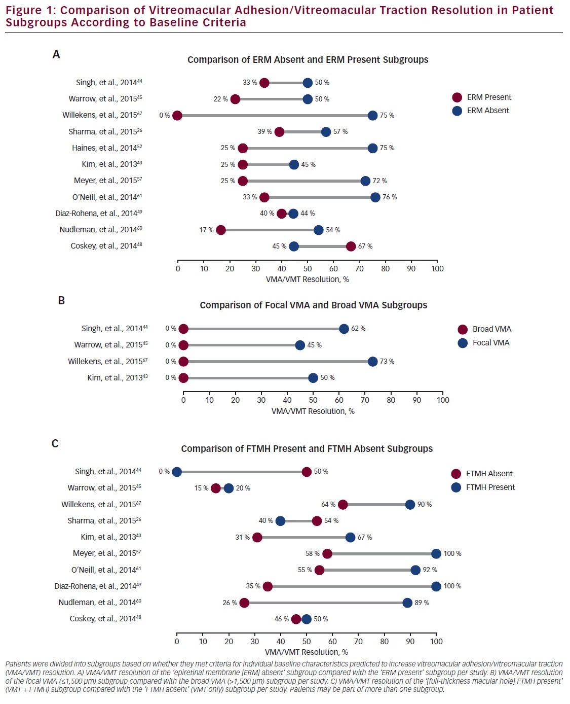
the overall average, emphasising the importance of appropriate patient selection. The comparisons between the ‘ERM absent,’ ‘focal VMA’ and ‘FTMH present’ subgroups and their corresponding non-criteriamatching counterparts are shown in Figure 1. In the majority of instances, the baseline predictor subgroup had higher VMA/VMT resolution. For instance, Sharma et al. reported an overall VMA/VMT resolution rate of 50 % in a study of 58 eyes, which increased to 57 % in the ‘ERM absent’ subgroup but decreased to 39 % in the ‘ERM present’ subgroup (see Figure 1A).26 Singh et al. reported a VMA/VMT resolution rate of 50 % for the ‘ERM absent’ subgroup versus 33 % for the ‘ERM present’ subgroup in a study with an overall resolution rate of 47 %.44 In this study, the focal VMA subgroup showed a resolution of 62 % compared with 0 % for patients with broad VMA (see Figure 1B). Kim et al. reported a resolution rate for the ‘FTMH present’ subgroup as 67 %, but only 31 % for the ‘FTMH absent’ subgroup (see Figure 1C).43 Even studies with high overall VMT resolution rates showed subgroup favourability. For instance, Willekens et al. reported an overall VMT resolution rate of 71 %; this rate is exceeded in every subgroup (see Figure 1A–C), and eyes meeting three criteria showed a resolution rate of 84 %.67

Overall, 100 % (4/4) of studies reporting results for both focal and broad VMA patients showed that the focal VMA subgroup exceeded the broad VMA subgroup VMA/VMT resolution rate. Similarly, 91 % (10/11) of studies for the ‘ERM absent’ subgroup exceeded the VMA/ VMT resolution rate of the ‘ERM present’ subgroup. Likewise, 80 % (8/10) of studies reported that the ‘FTMH present’ subgroup exceeded the ‘FTMH absent’ subgroup in terms of the VMA/VMT resolution rate. These results confirm that patient selection influences the assessment of ocriplasmin efficacy for VMA/VMT resolution.

In addition to VMA/VMT resolution, the efficacy of ocriplasmin for FTMH closure was assessed. In the clinical trials, closure rates were 40.6 % for ocriplasmin-treated versus 10.6 % for vehicle-treated patients at day 28, and remained high at the end of the study (40.6 % versus 17.0 %, respectively).40,41 Among the 26 studies, 22 reported FTMH closure rates (see Table 1).1,26,43–45,47–49,52,54–65,67 Of these, nine reported FTMH closure rates of 40 % or greater,43,44,52,54–56,60,64,67 and seven were 50 % or greater.43,44,52,54–56,60 FTMH sizes were not consistently reported in these studies, and closure rates have been shown to vary following

ocriplasmin treatment according to FTMH width at baseline.15,40,42 In addition, the duration of a macular hole has been shown to have an impact on the likelihood of spontaneous closure.28 FTMH closure may be directly related to VMT release; in one study, 81.8 % (9/11) of eyes that did not have hole closure had persistent VMT.26 This suggests that the mechanism of ocriplasmin-induced closure of FTMH may rely on prior VMT release.

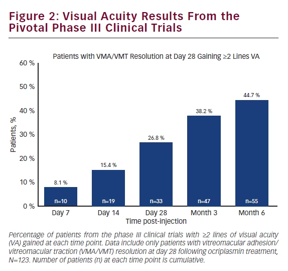
Analysis of the results from the two pivotal phase III MIVI-TRUST clinical trials showed that patients receiving ocriplasmin with VMA/VMT release by day 28 (primary endpoint) continued to show improvements in VA over time. By post-injection day 7, 8.1 % of these patients showed a ≥2 line improvement in VA; this number increased to 26.8 % at day 28 and 44.7 % at post-injection month 6 (see Figure 2).

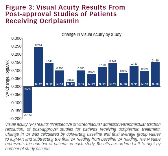
Similar to the clinical trials, many post-approval studies also tracked VA results; out of 26 published studies, 18 reported VA outcomes. Of these, 13 reported study group averages before and after ocriplasmin treatment, with the vast majority (12) reporting mean VA improvement26,44,45,48,50,53–57,59,62 and one reporting VA decline (see Figure 3).63 Study follow-up times ranged from 19 days to 8.7 months. Three of 18 studies reported VA improvement in a subset of patients; two studies showed VA improvement in patients who achieved VMA/ VMT release,1,60 and one study showed patient VA improvement following FTMH closure, but not for patients without FTMH closure.58 In contrast to the group averages, two of 18 studies reported VA outcomes on an individual basis. For instance, Kim et al. reported VA improvement in 52.6 % (10/19) patients,43 whereas Knudsen et al. reported improvement in 12.5 % (1/8) of patients.38

In addition to the 26 published studies, 20 case reports of patients receiving ocriplasmin treatment were published (in 16 articles) from 2013 to 2015.68–83 Case reports provide unique insight into ocriplasmin efficacy. Unlike studies, which have larger patient populations but present VA averages, case reports typically provide many more interim VA points to show progress over time for a single patient. Four cases were excluded due to patients receiving alternative treatment intervention prior to the final VA reading, and one case was excluded because the ocriplasmin treatment was unrelated to VMA/VMT, leaving
15 cases with analysable VA data.68–76,79,80,82,83 Ten of these 15 (66.7 %) reported improved VA results.70,73–76,79,80,83 Patients showing improvement were followed anywhere from 28 days to 15 months. Only three of the five cases not reporting VA improvement provided a follow-up length longer than 9 days. Of these, the longest reported follow-up period was 4 months.71,72,82 Importantly, of the 10 cases reporting VA improvement at the final follow-up, six (60 %) showed interim VA decline.73–75,79,80,83 This suggests that efficacy regarding VA improvement is closely related to the length of time patients are followed and reported on.

Discussion
Three years following the pivotal clinical trials and subsequent FDA approval of ocriplasmin, a more complete picture of ocriplasmin efficacy is beginning to emerge. Real-world efficacy results for ocriplasmin are now available for VMA/VMT resolution, FTMH closure and VA outcomes. Regarding VMA/VMT resolution, 92.3 % (24/26) of studies have now reported efficacy rates higher than the pivotal phase III clinical trials; 88 % (23/26) of these studies reported efficacy results higher than 40 %. Subgroup analysis showed that the baseline predictive factors derived from the clinical trials were validated in real-world studies; all studies (26/26) showed at least one subgroup with higher VMT resolution rates than the overall study average, and the great majority of subgroups consisting of baseline predictive factors outperformed their corresponding subgroups without those factors. Several studies showed further efficacy increases in patients with multiple baseline predictive factors.

Results regarding FTMH closure were more varied, but showed that 41 % (9/22) of studies reported results equal to or greater than those from the clinical trials. Further consideration of the baseline characteristics associated with higher rates of FTMH closure following treatment with ocriplasmin may be needed to define predictive factors. For VA improvement, a majority of studies (66.7 %, 12/18) reported average VA improvement in the study group, with five studies reporting VA improvement in a proportion of patients. The minimum follow-up time for these studies was 28 days. Similarly, 66.7 % (10/15) of case studies reported VA improvement following ocriplasmin treatment despite multiple patients experiencing a temporary VA decrease. Case reports provide valuable insights but need to be analysed collectively. Efficacy is determined by the endpoint; follow-up times varied widely, and VA can improve for more than 1 year. Moreover, short-term VA loss is usually recovered, which may not be captured in shorter followup periods. In addition, bias may exist to report n=1 cases showing unusual clinical circumstances, involving worsening VA outcomes.

Currently, a true direct comparison of ocriplasmin versus vitrectomy would require a randomised controlled clinical trial. However, ocriplasmin may provide an alternative therapeutic option to vitrectomy for suitable candidates. Although vitrectomy is highly effective, patients do experience a treatment burden of being unable to participate in daily activities for up to several weeks.36 Ocriplasmin may additionally provide an alternative to vitrectomy in patients not considered current vitrectomy candidates, such as patients with relatively good VA but severe symptoms.14 In one study, eyes with higher pretreatment BCVA had higher rates of VMT release following ocriplasmin treatment than eyes with worse pretreatment BCVA.26 This suggests that early treatment, before significant VA decline, may result in better VMA/ VMT resolution outcomes. Ocriplasmin intervention that improved visual function was shown to improve quality of life as assessed by the National Eye Institute 25-Item Visual Function Questionnaire (VFQ-25).84

The full clinical picture of ocriplasmin efficacy will be determined through continued and consistent reporting and data analysis. Accurate analysis, including study averages, as well as subgroups and collective case reports, will help further identify ideal candidates for ocriplasmin treatment.

Article Information:
Disclosure

Baruch D Kuppermann has received clinical research funding from Alcon, Allegro, Allergan, Genentech, GSK, Neurotech, Ophthotech, Regeneron and
ThromboGenics, and has been a consultant to AcuFocus, Aerpio, Alcon, Alimera, Allegro, Allergan, Ampio, Genentech, Neurotech, Novartis, Ophthotech, Regeneron,
SecondSight, STAAR Surgical, Teva and ThromboGenics.

Correspondence

Baruch D Kuppermann, Professor of Ophthalmology and Biomedical Engineering; Chief, Retina Service; Vice Chair, Academic Affairs, Gavin Herbert Eye
Institute, University of California, Irvine, Irvine, CA 92697, US. E: bdkupper@uci.edu

Access

This article is published under the Creative Commons Attribution Noncommercial License, which permits any noncommercial use, distribution, adaptation,
and reproduction provided the original author(s) and source are given appropriate credit.

Acknowledgements

Editorial assistance was provided by Meridius Health Communications, Inc. and funded by ThromboGenics

Received

2015-10-17T00:00:00

References

1. Chatziralli I, Theodossiadis G, Parikakis E, et al., Real-life experience after intravitreal ocriplasmin for vitreomacular traction and macular hole: a spectral-domain optical coherence tomography prospective study, Graefes Arch Clin Exp Ophthalmol, 2015 [Epub ahead of print].
2. García-Layana A, García-Arumí J, Ruiz-Moreno JM, et al., A review of current management of vitreomacular traction and macular hole, J Ophthalmol, 2015;2015:809640.
3. Steel DH, Lotery AJ, Idiopathic vitreomacular traction and macular hole: a comprehensive review of pathophysiology, diagnosis, and treatment, Eye (Lond), 2013;27(Suppl. 1):S1–21.
4. Yonemoto J, Noda Y, Masuhara N, Ohno S, Age of onset of posterior vitreous detachment, Curr Opin Ophthalmol, 1996;7:73–6.
5. Stalmans P, Duker JS, Kaiser PK, et al., Oct-based interpretation of the vitreomacular interface and indications for pharmacologic vitreolysis, Retina, 2013;33:2003–11.
6. Shao L, Wei W, Vitreomacular traction syndrome, Chin Med J (Engl), 2014;127:1566–71.
7. John VJ, Flynn HW, Jr, Smiddy WE, et al., Clinical course of vitreomacular adhesion managed by initial observation, Retina, 2014;34:442–6.
8. Johnson MW, Posterior vitreous detachment: evolution and complications of its early stages, Am J Ophthalmol, 2010;149:371–82 e371.
9. Johnson MW, Van Newkirk MR, Meyer KA, Perifoveal vitreous detachment is the primary pathogenic event in idiopathic macular hole formation, Arch Ophthalmol, 2001;119:215–22.
10. Schumann RG, Schaumberger MM, Rohleder M, et al., Ultrastructure of the vitreomacular interface in full-thickness idiopathic macular holes: a consecutive analysis of 100 cases, Am J Ophthalmol, 2006;141:1112–9.
11. Smiddy WE, Flynn HW, Jr, Pathogenesis of macular holes and therapeutic implications, Am J Ophthalmol, 2004;137:525–37.
12. Spaide RF, Closure of an outer lamellar macular hole by vitrectomy: hypothesis for one mechanism of macular hole formation, Retina, 2000;20:587–90.
13. Duker JS, Kaiser PK, Binder S, et al., The International Vitreomacular Traction Study Group classification of vitreomacular adhesion, traction, and macular hole, Ophthalmology, 2013;120:2611–9.
14. Song SJ, Smiddy WE, Ocriplasmin for symptomatic vitreomacular adhesion: an evidence-based review of its potential, Core Evid, 2014;9:51–9.
15. Stefanini FR, Maia M, Falabella P, et al., Profile of ocriplasmin and its potential in the treatment of vitreomacular adhesion, Clin Ophthalmol, 2014;8:847–56.
16. Uchino E, Uemura A, Ohba N, Initial stages of posterior vitreous detachment in healthy eyes of older persons evaluated by optical coherence tomography, Arch Ophthalmol, 2001;119:1475–9.
17. Almeida DR, Chin EK, Rahim K, et al., Factors associated with spontaneous release of vitreomacular traction, Retina, 2015;35:492–7.
18. Codenotti M, Iuliano L, Fogliato G, et al., A novel spectraldomain optical coherence tomography model to estimate changes in vitreomacular traction syndrome, Graefes Arch Clin Exp Ophthalmol, 2014;252:1729–35.
19. Dimopoulos S, Bartz-Schmidt KU, Gelisken F, et al., Rate and timing of spontaneous resolution in a vitreomacular traction group: should the role of watchful waiting be re-evaluated as an alternative to ocriplasmin therapy?, Br J Ophthalmol, 2015;99:350–3.
20. Theodossiadis G, Petrou P, Eleftheriadou M, et al., Focal vitreomacular traction: a prospective study of the evolution to macular hole: the mathematical approach, Eye (Lond), 2014;28:1452–60.
21. Theodossiadis GP, Chatziralli IP, Sergentanis TN, et al., Evolution of vitreomacular adhesion to acute vitreofoveal separation with special emphasis on a traction-induced foveal pathology, A prospective study of spectral-domain optical coherence tomography, Graefes Arch Clin Exp Ophthalmol, 2015;253:1425–35.
22. Theodossiadis GP, Grigoropoulos VG, Theodoropoulou S, et al., Spontaneous resolution of vitreomacular traction demonstrated by spectral-domain optical coherence tomography, Am J Ophthalmol, 2014;157:842–51 e841.
23. Zhang Z, Dong F, Zhao C, et al., Natural course of vitreomacular traction syndrome observed by spectraldomain optical coherence tomography, Can J Ophthalmol, 2015;50:172–9.
24. Hikichi T, Yoshida A, Trempe CL, Course of vitreomacular traction syndrome, Am J Ophthalmol, 1995;119:55–61.
25. Sonmez K, Capone A, Jr, Trese MT, Williams GA, Vitreomacular traction syndrome: impact of anatomical configuration on anatomical and visual outcomes, Retina, 2008;28:1207–14.
26. Sharma P, Juhn A, Houston SK, et al., Efficacy of intravitreal ocriplasmin on vitreomacular traction and full-thickness macular holes, Am J Ophthalmol, 2015;159:861–7 e862.
27. Kim JW, Freeman WR, Azen SP, et al., Prospective randomized trial of vitrectomy or observation for stage 2 macular holes, Vitrectomy for Macular Hole Study Group, Am J Ophthalmol, 1996;121:605–14.
28. Jaycock PD, Bunce C, Xing W, et al., Outcomes of macular hole surgery: implications for surgical management and clinical governance, Eye (Lond), 2005;19:879–84.
29. Margherio RR, Trese MT, Margherio AR, Cartright K, Surgical management of vitreomacular traction syndromes, Ophthalmology, 1989;96:1437–45.
30. Smiddy WE, Michels RG, Glaser BM, deBustros S, Vitrectomy for macular traction caused by incomplete vitreous separation, Arch Ophthalmol, 1988;106:624–8.
31. Mester V, Kuhn F, Internal limiting membrane removal in the management of full-thickness macular holes, Am J Ophthalmol, 2000;129:769–77.
32. Meng Q, Zhang S, Ling Y, et al., Long-term anatomic and visual outcomes of initially closed macular holes, Am J Ophthalmol, 2011;151:896–900 e892.
33. Benson WE, Cruickshanks KC, Fong DS, et al., Surgical management of macular holes: a report by the American Academy of Ophthalmology, Ophthalmology, 2001;108:1328–35.
34. Christensen UC, Kroyer K, Sander B, et al., Value of internal limiting membrane peeling in surgery for idiopathic macular hole stage 2 and 3: a randomised clinical trial, Br J Ophthalmol, 2009;93:1005–15.
35. Jackson TL, Nicod E, Angelis A, et al., Pars plana vitrectomy for vitreomacular traction syndrome: a systematic review and metaanalysis of safety and efficacy, Retina, 2013;33:2012–7.
36. Ocriplasmin (Jetrea) (125 mcg Intravitreal Injection): For the Treatment of Symptomatic Vitreomacular Adhesion, Ottawa (ON); 2014.
37. Johnson MW, How should we release vitreomacular traction: surgically, pharmacologically, or pneumatically? Am J Ophthalmol, 2013;155:203–5 e201.
38. Knudsen VM, Kozak I, A retrospective study of a single practice use of ocriplasmin in the treatment of vitreomacular traction, Saudi J Ophthalmol, 2014;28:139–44.
39. Chen W, Mo W, Sun K, et al., Microplasmin degrades fibronectin and laminin at vitreoretinal interface andouter retina during enzymatic vitrectomy, Curr Eye Res, 2009;34:1057.64.
40. Stalmans P, Benz MS, Gandorfer A, et al., Enzymatic vitreolysis with ocriplasmin for vitreomacular traction and macular holes, N Engl J Med, 2012;367:606.15.
41. Syed YY, Dhillon S, Ocriplasmin: a review of its use in patients with symptomatic vitreomacular adhesion, Drugs, 2013;73:1617.25.
42. Haller JA, Stalmans P, Benz MS, et al., Efficacy of intravitreal ocriplasmin for treatment of vitreomacular adhesion: subgroup analyses from two randomized trials, Ophthalmology, 2015;122:117.22.
43. Kim BT, Schwartz SG, Smiddy WE, et al., Initial outcomes following intravitreal ocriplasmin for treatment of symptomatic vitreomacular adhesion, Ophthalmic Surg Lasers Imaging Retina, 2013;44:334.43.
44. Singh RP, Li A, Bedi R, et al., Anatomical and visual outcomes following ocriplasmin treatment for symptomatic vitreomacular traction syndrome, Br J Ophthalmol, 2014;98:356.60.
45. Warrow DJ, Lai MM, Patel A, et al., Treatment outcomes and spectral-domain optical coherence tomography findings of eyes with symptomatic vitreomacular adhesion treated with intravitreal ocriplasmin, Am J Ophthalmol, 2015;159:20.30 e21.
46. Khan MA, Haller JA, Clinical management of vitreomacular traction, Curr Opin Ophthalmol, 2015;26:143.8.
47. Chin EK, Almeida DR, Sohn EH, et al., Incomplete vitreomacular traction release using intravitreal ocriplasmin, Case Rep Ophthalmol, 2014;5:455.62.
48. Coskey A, Brown DM, Hooten C, et al., Ocriplasmin for vitreomacular adhesion (VMA) in the clinical setting: rates of VMA release, development of macular hole, and visual outcomes. Presented at: The Association for Research in Vision and Ophthalmology (ARVO) Annual Meeting; 4.8 May 2014; Orlando, FL.
49. Diaz-Rohena R, Flores-Sanchez B, Ocriplasmin in clinical practice. Presented at: Pan-American Retina and Vitreous Society (SPRV) Meeting; 11.14 June 2014; San Juan, Puerto Rico.
50. Fineman MS, For suitable sVMA patients, ocriplasmin has eno down sidef, Ocular Surgery News, Improving Patient Outcomes in Symptomatic VMA (supplement), 2013;7.10.
51. Hager A, Seibel I, Riechardt A, et al., Does ocriplasmin affect the RPE-photoreceptor adhesion in macular holes? Br J Ophthalmol, 2015;99:635.8.
52. Haines N, Copeland V, Kurup S, et al., Outcomes of treatment with intravitreal ocriplasmin for vitreomacular traction syndrome and macular holes at the Wake Forest Eye Center. Presented at: American Society of Retina Specialists (ASRS) Annual Meeting; 9.13 August 2014; San Diego, CA.
53. Itoh Y, Kaiser PK, Singh RP, et al., Assessment of retinal alterations after intravitreal ocriplasmin with spectraldomain optical coherence tomography, Ophthalmology, 2014;121:2506.7 e2502.
54. Khanani AM, Improving success with pharmacologic management of symptomatic VMA, Retina Today, 2015;January/February:48.50.
55. Lakhanpal RR, New injection gives the ewait and seef patient a chance for symptom relief, Ocular Surgery News, Improving Patient Outcomes in Symptomatic VMA (supplement), 2013;4.6.
56. Maier MM, Bonse S, Frank C, et al., Jetrea (ocriplasmin) as a treatment option for symptomatic vitreomacular traction with or without macular hole (.400 ƒÊm) . first clinical experience. Presented at: The Association for Research in Vision and Ophthalmology (ARVO) Annual Meeting; 4.8 May 2014; Orlando, FL.
57. Meyer JC, Shah GK, Blinder KJ, et al., Early evolution of the vitreomacular interface and clinical efficacy after ocriplasmin injection for symptomatic vitreomacular adhesion, Ophthalmic Surg Lasers Imaging Retina, 2015;46:209.16.
58. Miller JB, Kim LA, Wu DM, et al., Ocriplasmin for treatment of stage 2 macular holes: early clinical results, Ophthalmic Surg Lasers Imaging Retina, 2014;45:293.7.
59. Nasir M, Pieramici D, Castellarin A, Intravitreal ocriplasmin for vitreomacular adhesion syndrome: initial experience of multiphysician retina practice. Presented at: American Society of Retina Specialists (ASRS) Annual Meeting; 24.28 August 2013; Toronto, Canada.
60. Nudleman E, Franklin MS, Ruby AJ, Wolfe JD, Ocriplasmin for vitreomacular adhesion: aftermarket experience and findings. Presented at: American Society of Retina Specialists (ASRS) Annual Meeting; 9.13 August 2014; San Diego, CA.
61. OfNeill BP, Shah AP, Coney JM, Resolution of vitreomacular traction using ocriplasmin (Jetrea) and its potential role in diabetic macular edema. Presented at: The Association for Research in Vision and Ophthalmology (ARVO) Annual Meeting; 4.8 May 2014; Orlando, FL.
62. Quezada-Ruiz C, Pieramici DJ, Nasir M, et al., Outer retina reflectivity changes on sd-oct after intravitreal ocriplasmin for vitreomacular traction and macular hole, Retina, 2015;35:1144.50.
63. Reiss B, Smithen L, Mansour S, Transient vision loss after ocriplasmin injection, Retina, 2015;35:1107.10.
64. Roth D, Feng HL, Modi KK, et al., Predictors of success with intravitreal ocriplasmin in the treatment of symptomatic vitreomacular adhesion. Presented at: The Association for Research in Vision and Ophthalmology (ARVO) Annual Meeting; 4.8 May 2014; Orlando, FL.
65. Steel DH, Sandinha MT, White K, The Plane of Vitreoretinal Separation and Results of Vitrectomy Surgery in Patients Given Ocriplasmin for Idiopathic Macular Hole, Invest Ophthalmol Vis Sci, 2015;56:4038.44.
66. Torres W, Initial outcomes of intravitreal ocriplasmin (Jetrea) at Sarasota Retina Institute. Presented at: 28th Annual Sarasota Vitreo-Retinal Update Course; 13.15 February 2014; Sarasota, FL, US.
67. Willekens K, Abegao Pinto L, Vandewalle E, et al., Improved efficacy of ocriplasmin for vitreomacular traction release and transient changes in optic disk morphology, Retina, 2015;35:1135.43.
68. Barteselli G, Carini E, Invernizzi A, et al., Early panretinal abnormalities on fundus autofluorescence and spectral domain optical coherence tomography after intravitreal ocriplasmin, Acta Ophthalmol, 2015 [Epub ahead of print].
69. Casswell E, Fernandez-Sanz G, Mitry D, et al., Macular Hole Progression following Ocriplasmin Intravitreal Injection, Case Rep Ophthalmol Med, 2014;2014:403461.
70. Chod RB, Goodrich C, Saxena S, Akduman L, Lamellar macular hole after intravitreal ocriplasmin injection, BMJ Case Rep, 2015;2015.
71. DaCosta J, Younis S, Transient visual loss and delayed resolution of vitreomacular traction after intravitreal ocriplasmin, Drug Healthc Patient Saf, 2014;6:175.8.
72. Fahim AT, Khan NW, Johnson MW, Acute panretinal structural and functional abnormalities after intravitreous ocriplasmin injection, JAMA Ophthalmol, 2014;132:484.6.
73. Freund KB, Shah SA, Shah VP, Correlation of transient vision loss with outer retinal disruption following intravitreal ocriplasmin, Eye (Lond), 2013;27:773.4.
74. Han IC, Scott AW, Sterile endophthalmitis after intravitreal ocriplasmin injection: report of a single case, Retin Cases Brief Rep, 2015;9:242.4.
75. Jeng KW, Baumal CR, Witkin AJ, et al., Incomplete release of vitreomacular attachments after intravitreal ocriplasmin, Ophthalmic Surg Lasers Imaging Retina, 2015;46:271.4.
76. Katz RS, Bilateral vitreomacular traction resolved with a single intravitreal ocriplasmin injection, Ophthalmic Surg Lasers Imaging Retina, 2014;45:239.42.
77. Keller J, Haynes RJ, Zonular Dehiscence at the Time of Combined Vitrectomy and Cataract Surgery After Intravitreal Ocriplasmin Injection, JAMA Ophthalmol, 2015;133:1091.2.
78. Patel A, Morse L, Ocriplasmin for foveal schisis in x-linked retinoschisis, Retin Cases Brief Rep, 2015;9:248.51.
79. Quezada-Ruiz C, Pieramici DJ, Nasir M, et al., Severe acute vision loss, dyschromatopsia, and changes in the ellipsoid zone on sd-oct associated with intravitreal ocriplasmin injection, Retin Cases Brief Rep, 2015;9:145.8.
80. Reiss B, Smithen L, Mansour S, Acute vision loss after ocriplasmin use, Retin Cases Brief Rep, 2015;9:168.9.
81. Silva RA, Moshfeghi DM, Leng T, Retinal breaks due to intravitreal ocriplasmin, Clin Ophthalmol, 2014;8:1591.4.
82. Thanos A, Hernandez-Siman J, Marra KV, Arroyo JG, Reversible vision loss and outer retinal abnormalities after intravitreal ocriplasmin injection, Retin Cases Brief Rep, 2014;8:330.2.
83. Tibbetts MD, Reichel E, Witkin AJ, Vision loss after intravitreal ocriplasmin: correlation of spectral-domain optical coherence tomography and electroretinography, JAMA Ophthalmol, 2014;132:487.90.
84. Hirneiss C, Neubauer AS, Gass CA, et al., Visual quality of life after macular hole surgery: outcome and predictive factors, Br J Ophthalmol, 2007;91:481.4.

Further Resources

Share this Article
Related Content In Retina/Vitreous
  • Copied to clipboard!
    accredited arrow-down-editablearrow-downarrow_leftarrow-right-bluearrow-right-dark-bluearrow-right-greenarrow-right-greyarrow-right-orangearrow-right-whitearrow-right-bluearrow-up-orangeavatarcalendarchevron-down consultant-pathologist-nurseconsultant-pathologistcrosscrossdownloademailexclaimationfeedbackfiltergraph-arrowinterviewslinkmdt_iconmenumore_dots nurse-consultantpadlock patient-advocate-pathologistpatient-consultantpatientperson pharmacist-nurseplay_buttonplay-colour-tmcplay-colourAsset 1podcastprinter scenerysearch share single-doctor social_facebooksocial_googleplussocial_instagramsocial_linkedin_altsocial_linkedin_altsocial_pinterestlogo-twitter-glyph-32social_youtubeshape-star (1)tick-bluetick-orangetick-red tick-whiteticktimetranscriptup-arrowwebinar Sponsored Department Location NEW TMM Corporate Services Icons-07NEW TMM Corporate Services Icons-08NEW TMM Corporate Services Icons-09NEW TMM Corporate Services Icons-10NEW TMM Corporate Services Icons-11NEW TMM Corporate Services Icons-12Salary £ TMM-Corp-Site-Icons-01TMM-Corp-Site-Icons-02TMM-Corp-Site-Icons-03TMM-Corp-Site-Icons-04TMM-Corp-Site-Icons-05TMM-Corp-Site-Icons-06TMM-Corp-Site-Icons-07TMM-Corp-Site-Icons-08TMM-Corp-Site-Icons-09TMM-Corp-Site-Icons-10TMM-Corp-Site-Icons-11TMM-Corp-Site-Icons-12TMM-Corp-Site-Icons-13TMM-Corp-Site-Icons-14TMM-Corp-Site-Icons-15TMM-Corp-Site-Icons-16TMM-Corp-Site-Icons-17TMM-Corp-Site-Icons-18TMM-Corp-Site-Icons-19TMM-Corp-Site-Icons-20TMM-Corp-Site-Icons-21TMM-Corp-Site-Icons-22TMM-Corp-Site-Icons-23TMM-Corp-Site-Icons-24TMM-Corp-Site-Icons-25TMM-Corp-Site-Icons-26TMM-Corp-Site-Icons-27TMM-Corp-Site-Icons-28TMM-Corp-Site-Icons-29TMM-Corp-Site-Icons-30TMM-Corp-Site-Icons-31TMM-Corp-Site-Icons-32TMM-Corp-Site-Icons-33TMM-Corp-Site-Icons-34TMM-Corp-Site-Icons-35TMM-Corp-Site-Icons-36TMM-Corp-Site-Icons-37TMM-Corp-Site-Icons-38TMM-Corp-Site-Icons-39TMM-Corp-Site-Icons-40TMM-Corp-Site-Icons-41TMM-Corp-Site-Icons-42TMM-Corp-Site-Icons-43TMM-Corp-Site-Icons-44TMM-Corp-Site-Icons-45TMM-Corp-Site-Icons-46TMM-Corp-Site-Icons-47TMM-Corp-Site-Icons-48TMM-Corp-Site-Icons-49TMM-Corp-Site-Icons-50TMM-Corp-Site-Icons-51TMM-Corp-Site-Icons-52TMM-Corp-Site-Icons-53TMM-Corp-Site-Icons-54TMM-Corp-Site-Icons-55TMM-Corp-Site-Icons-56TMM-Corp-Site-Icons-57TMM-Corp-Site-Icons-58TMM-Corp-Site-Icons-59TMM-Corp-Site-Icons-60TMM-Corp-Site-Icons-61TMM-Corp-Site-Icons-62TMM-Corp-Site-Icons-63TMM-Corp-Site-Icons-64TMM-Corp-Site-Icons-65TMM-Corp-Site-Icons-66TMM-Corp-Site-Icons-67TMM-Corp-Site-Icons-68TMM-Corp-Site-Icons-69TMM-Corp-Site-Icons-70TMM-Corp-Site-Icons-71TMM-Corp-Site-Icons-72